文献精选
Denise Türk1 iD Nina Scherer1 & Dominik Selzer1 & Christiane Dings1 & Nina Hanke1 & Robert Dallmann2 iD Matthias Schwab3,4,5 & Peter Timmins 6 iD Valerie Nock7 & Thorsten Lehr1 iD
Received: 22 November 2022 /Accepted: 31 January 2023 © The Author(s) 2023
Abstract
Aims/hypothesis The objective was to investigate if metformin pharmacokinetics is modulated by time-of-day in humans using empirical and mechanistic pharmacokinetic modelling techniques on a large clinical dataset. This study also aimed to generate and test hypotheses on the underlying mechanisms, including evidence for chronotype-dependent interindividual differences in metformin plasma and efficacy-related tissue concentrations.
Methods A large clinical dataset consisting of individual metformin plasma and urine measurements was analysed using a newly developed empirical pharmacokinetic model. Causes of daily variation of metformin pharmacokinetics and interindividual variability were further investigated by a literature-informed mechanistic modelling analysis.
Results A significant effect of time-of-day on metformin pharmacokinetics was found. Daily rhythms of gastrointestinal, hepatic and renal processes are described in the literature, possibly affecting drug pharmacokinetics. Observed metformin plasma levels were best described by a combination of a rhythm in GFR, renal plasma flow (RPF) and organic cation transporter (OCT) 2 activity. Furthermore, the large interindividual differences in measured metformin concentrations were best explained by individual chronotypes affecting metformin clearance, with impact on plasma and tissue concentrations that may have implications for metformin efficacy.
Conclusions/interpretation Metformin’s pharmacology significantly depends on time-of-day in humans, determined with the help of empirical and mechanistic pharmacokinetic modelling, and rhythmic GFR, RPF and OCT2 were found to govern intraday variation. Interindividual variation was found to be partly dependent on individual chronotype, suggesting diurnal preference as an interesting, but so-far underappreciated, topic with regard to future personalised chronomodulated therapy in people with type 2 diabetes.
Keywords Chronopharmacology . Empirical modelling . Mechanistic modelling . Metformin . Pharmacokinetics . Renal excretion . Transporter
Thorsten Lehr
该Email地址已收到反垃圾邮件插件保护。要显示它您需要在浏览器中启用JavaScript。
1 Clinical Pharmacy, Saarland University, Saarbrücken, Germany
2 Division of Biomedical Sciences, Warwick Medical School, University of Warwick, Coventry, UK
3 Dr. Margarete Fischer-Bosch-Institute of Clinical Pharmacology, Stuttgart, Germany
4 Departments of Clinical Pharmacology, Pharmacy and Biochemistry, University of Tübingen, Tübingen, Germany
5 Cluster of Excellence iFIT (EXC2180) ‘Image-guided and Functionally Instructed Tumor Therapies’, University of Tübingen, Tübingen, Germany
6 Department of Pharmacy, University of Huddersfield, Huddersfield, UK
7 Boehringer Ingelheim Pharma GmbH & Co. KG, Biberach, Germany
Georgios E. Papanikolaou 1,*, Georgios Gousios 2 and Niels A. J. Cremers 3,4,*iD
1 GP Plastic Surgery Private Practice, P. Dagkli 1, 45444 Ioannina, Greece
2 PharmaLife, I. Vilara 40, 45444 Ioannina, Greece
3 Department of Gynecology and Obstetrics, Maastricht University Medical Centre, P. Debyelaan 25, 6229 HX Maastricht, The Netherlands
4 Triticum Exploitatie BV, Sleperweg 44, 6222 NK Maastricht, The Netherlands
*Correspondence: 该Email地址已收到反垃圾邮件插件保护。要显示它您需要在浏览器中启用JavaScript。 (G.E.P.); 该Email地址已收到反垃圾邮件插件保护。要显示它您需要在浏览器中启用JavaScript。 or 该Email地址已收到反垃圾邮件插件保护。要显示它您需要在浏览器中启用JavaScript。 (N.A.J.C.); Tel.: +30-2651-607063 (G.E.P.); +31-43-325-1773 (N.A.J.C.)
Abstract: Management of locally infected heel-pressure ulcers (HPUs) remains challenging, and given the increasing occurrence of infections resistant to antibiotic therapy and patients’ unwillingness to surgery, innovative and effective approaches must be considered. Medical-grade honey (MGH) could be an alternative therapeutic approach due to its broad-spectrum antimicrobial activity and healing properties. This study aimed to present the high effectiveness and safety of MGH for the conservative treatment of clinically infected HPUs. In this case series, we have prospectively studied nine patients with local signs of infected HPUs. In all cases, HPUs persisted for more than 4 weeks, and previous treatments with topical antibiotics or antiseptic products were ineffective. All patients were at high-risk to develop HPU infection due to their advanced age (median age of 86 years), several comorbidities, and permanent immobility. All wounds were treated with MGH products (L-Mesitran), leading to infection resolution within 3–4 weeks and complete wound healing without complication. Considering the failure of previous treatments and the chronic nature of the wounds, MGH was an effective treatment. MGH-based products are clinically and cost-effective for treating hard-to-heal pressure ulcers such as HPUs. Thus, MGH can be recommended as an alternative or complementary therapy in wound healing.
Keywords: medical-grade honey; heel-pressure ulcers; infection; antibiotic resistance; wounds; wound healing
Rianneke de Ritter1,2 ● Simone J. S. Sep1,2,3 & Marleen M. J. van Greevenbroek1,2 & Yvo H. A. M. Kusters1,2 & Rimke C. Vos4,5 ● Michiel L. Bots4 ● M. Eline Kooi2,6 ● Pieter C. Dagnelie1,2 ● Simone J. P. M. Eussen2,7,8 ● Miranda T. Schram1,2,9,10 ● Annemarie Koster8,11 ● Martijn C. G. Brouwers 1,2 ● Niels M. R. van der Sangen12 ● Sanne A. E. Peters4,13 ● Carla J. H. van der Kallen1,2 ● Coen D. A. Stehouwer1,2
Received: 27 May 2022 / Accepted: 22 November 2022 / Published online: 20 February 2023 © The Author(s) 2023
Abstract
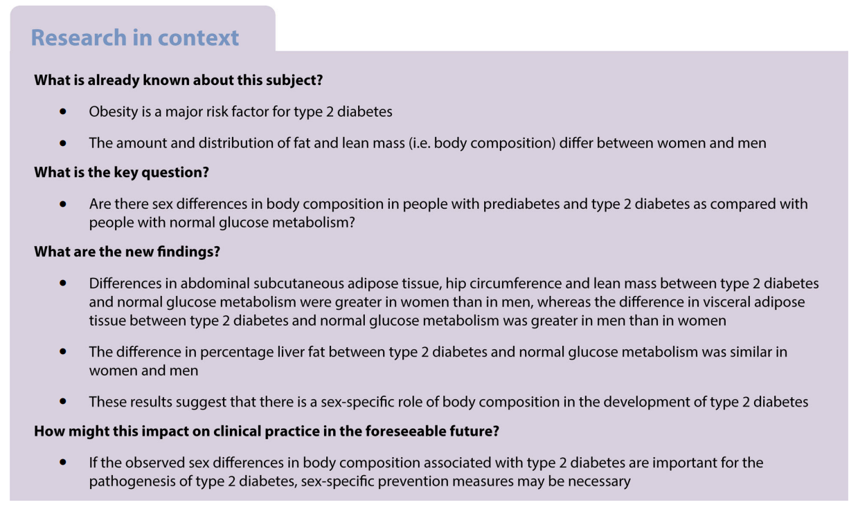
Aims/hypothesis Obesity is a major risk factor for type 2 diabetes. However, body composition differs between women and men. In this study we investigate the association between diabetes status and body composition and whether this association is moderated by sex.
Methods In a population-based cohort study (n=7639; age 40–75 years, 50% women, 25% type 2 diabetes), we estimated the sex-specific associations, and differences therein, of prediabetes (i.e. impaired fasting glucose and/or impaired glucose tolerance) and type 2 diabetes (reference: normal glucose metabolism [NGM]) with dual-energy x-ray absorptiometry (DEXA)- and MRIderived measures of body composition and with hip circumference. Sex differences were analysed using adjusted regression models with interaction terms of sex-by-diabetes status.
Results Compared with their NGM counterparts, both women and men with prediabetes and type 2 diabetes had more fat and lean mass and a greater hip circumference. The differences in subcutaneous adipose tissue, hip circumference and total and peripheral lean mass between type 2 diabetes and NGM were greater in women than men (women minus men [W–M] mean difference [95% CI]: 15.0 cm2 [1.5, 28.5], 3.2 cm [2.2, 4.1], 690 g [8, 1372] and 443 g [142, 744], respectively). The difference in visceral adipose tissue between type 2 diabetes and NGM was greater in men than women (W–M mean difference [95% CI]: −14.8 cm2 [−26.4, −3.1]). There was no sex difference in the percentage of liver fat between type 2 diabetes and NGM. The differences in measures of body composition between prediabetes and NGM were generally in the same direction, but were not significantly different between women and men.
Conclusions/interpretation This study indicates that there are sex differences in body composition associated with type 2 diabetes. The pathophysiological significance of these sex-associated differences requires further study.
Rianneke de Ritter
该Email地址已收到反垃圾邮件插件保护。要显示它您需要在浏览器中启用JavaScript。
1 Department of Internal Medicine, Maastricht University Medical Center+, Maastricht, the Netherlands
2 CARIM School for Cardiovascular Diseases, Maastricht University, Maastricht, the Netherlands
3 Adelante, Center of Expertise in Rehabilitation and Audiology, Hoensbroek, the Netherlands
4 Julius Center for Health Sciences and Primary Care, University Medical Center Utrecht, Utrecht University, Utrecht, the Netherlands
5 Leiden University Medical Center, Department of Public Health and Primary Care/LUMC-Campus, The Hague, the Netherlands
6 Department of Radiology and Nuclear Medicine, Maastricht University Medical Center+, Maastricht, the Netherlands
7 Department of Epidemiology, Maastricht University, Maastricht, the Netherlands
8 CAPHRI Care and Public Health Research Institute, Maastricht University, Maastricht, the Netherlands
9 Heart and Vascular Center, Maastricht University Medical Center+, Maastricht, the Netherlands
10 MHeNs School for Mental Health and Neuroscience, Maastricht University Medical Center+, Maastricht, the Netherlands
11 Department of Social Medicine, Maastricht University, Maastricht, the Netherlands
12 Department of Cardiology, Amsterdam UMC, University of Amsterdam, Amsterdam, the Netherlands
13 The George Institute for Global Health, Imperial College London, London, UK
Keywords Body composition . DEXA . Fat mass . Lean mass . Liver fat . MRI . Prediabetes . Sex differences . Type 2 diabetes . Women
Abbreviations
DEXA Dual-energy x-ray absorptiometry
GMR Geometric mean ratio
GMS Glucose metabolism status
NGM Normal glucose metabolism
SAT Subcutaneous adipose tissue
VAT Visceral adipose tissue
W–M Women minus men
Melanie Lloyd1,2 iD Jedidiah Morton1,2,3 ● Helena Teede2 ● Clara Marquina1 ● Dina Abushanab1 ● Dianna J. Magliano2,3 ● Emily J. Callander2 ● Zanfina Ademi1,2
Received: 25 August 2022 /Accepted: 31 January 2023 © The Author(s) 2023
Abstract
Aims/hypothesis The aim of this study was to determine the long-term cost-effectiveness and return on investment of implementing a structured lifestyle intervention to reduce excessive gestational weight gain and associated incidence of gestational diabetes mellitus (GDM) and type 2 diabetes mellitus.
Methods A decision-analytic Markov model was used to compare the health and cost-effectiveness outcomes for (1) a structured lifestyle intervention during pregnancy to prevent GDM and subsequent type 2 diabetes; and (2) current usual antenatal care. Life table modelling was used to capture type 2 diabetes morbidity, mortality and quality-adjusted life years over a lifetime horizon for all women giving birth in Australia. Costs incorporated both healthcare and societal perspectives. The intervention effect was derived from published meta-analyses. Deterministic and probabilistic sensitivity analyses were used to capture the impact of uncertainty in the model.
Results The model projected a 10% reduction in the number of women subsequently diagnosed with type 2 diabetes through implementation of the lifestyle intervention compared with current usual care. The total net incremental cost of intervention was approximately AU$70 million, and the cost savings from the reduction in costs of antenatal care for GDM, birth complications and type 2 diabetes management were approximately AU$85 million. The intervention was dominant (cost-saving) compared with usual care from a healthcare perspective, and returned AU$1.22 (95% CI 0.53, 2.13) per dollar invested. The results were robust to sensitivity analysis, and remained cost-saving or highly cost-effective in each of the scenarios explored.
Conclusions/interpretation This study demonstrates significant cost savings from implementation of a structured lifestyle intervention during pregnancy, due to a reduction in adverse health outcomes for women during both the perinatal period and over their lifetime.
Keywords Cost-effectiveness . Decision modelling . Dietary intervention . Epidemiology . Gestational diabetes mellitus . Life table modelling . Physical activity . Pregnancy . Type 2 diabetes mellitus
Zanfina Ademi
该Email地址已收到反垃圾邮件插件保护。要显示它您需要在浏览器中启用JavaScript。
1 Centre for Medicine Use and Safety, Faculty of Pharmacy and Pharmaceutical Sciences, Monash University, Melbourne, Australia
2 School of Public Health and Preventive Medicine, Monash University, Melbourne, Australia
3 Diabetes and Population Health, Baker Heart and Diabetes Institute, Melbourne, Australia
