文献精选


This article is excerpted from the Nature | Vol 635 | 21 November 2024 by Wound World.

Nusayhah Hudaa Gopee1,2,18, Elena Winheim3,18, Bayanne Olabi1,2,18, Chloe Admane1,3, April Rose Foster3 , Ni Huang3 , Rachel A. Botting1 , Fereshteh Torabi3 , Dinithi Sumanaweera3 , Anh Phuong Le4,5,6, Jin Kim4,5,6, Luca Verger7 , Emily Stephenson1,3, Diana Adão3 , Clarisse Ganier8 , Kelly Y. Gim4,5,6, Sara A. Serdy4,5,6, CiCi Deakin4,5,6, Issac Goh1,3, Lloyd Steele3 , Karl Annusver9 , Mohi-Uddin Miah1 , Win Min Tun1,3, Pejvak Moghimi3 , Kwasi Amoako Kwakwa3 , Tong Li3 , Daniela Basurto Lozada1 , Ben Rumney3 , Catherine L. Tudor3 , Kenny Roberts3 , Nana-Jane Chipampe3 , Keval Sidhpura1 , Justin Englebert1 , Laura Jardine1 , Gary Reynolds1 , Antony Rose1,3, Vicky Rowe3 , Sophie Pritchard3 , Ilaria Mulas3 , James Fletcher1 , Dorin-Mirel Popescu1 , Elizabeth Poyner1,2, Anna Dubois2 , Alyson Guy10, Andrew Filby1 , Steven Lisgo1 , Roger A. Barker11, Ian A. Glass12, Jong-Eun Park3 , Roser Vento-Tormo3 , Marina Tsvetomilova Nikolova13, Peng He3,14, John E. G. Lawrence3 , Josh Moore15, Stephane Ballereau3 , Christine B. Hale3 , Vijaya Shanmugiah3 , David Horsfall1 , Neil Rajan1,2, John A. McGrath16, Edel A. O’Toole17, Barbara Treutlein13, Omer Bayraktar3 , Maria Kasper9 , Fränze Progatzky7 , Pavel Mazin3 , Jiyoon Lee4,5,6, Laure Gambardella3 , Karl R. Koehler4,5,6,19 ✉, Sarah A. Teichmann3,19 ✉ & Muzlifah Haniffa1,2,3,19 ✉
https://doi.org/10.1038/s41586-024-08002-x
Received: 4 August 2023
Accepted: 28 August 2024
Published online: 16 October 2024
Open access
Check for updates
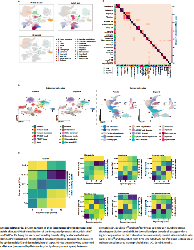
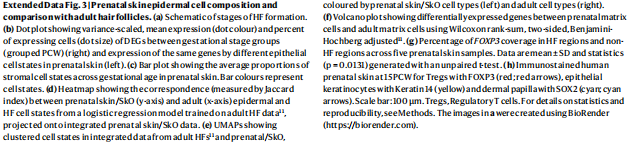
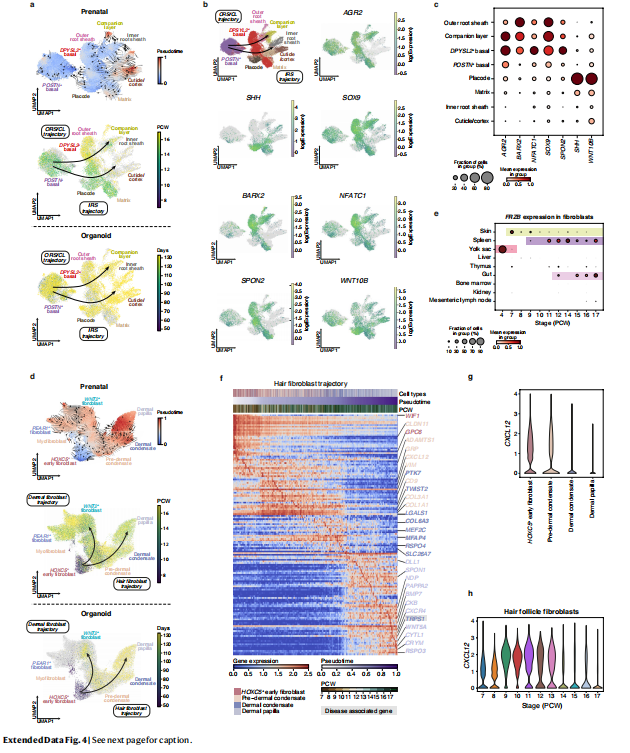
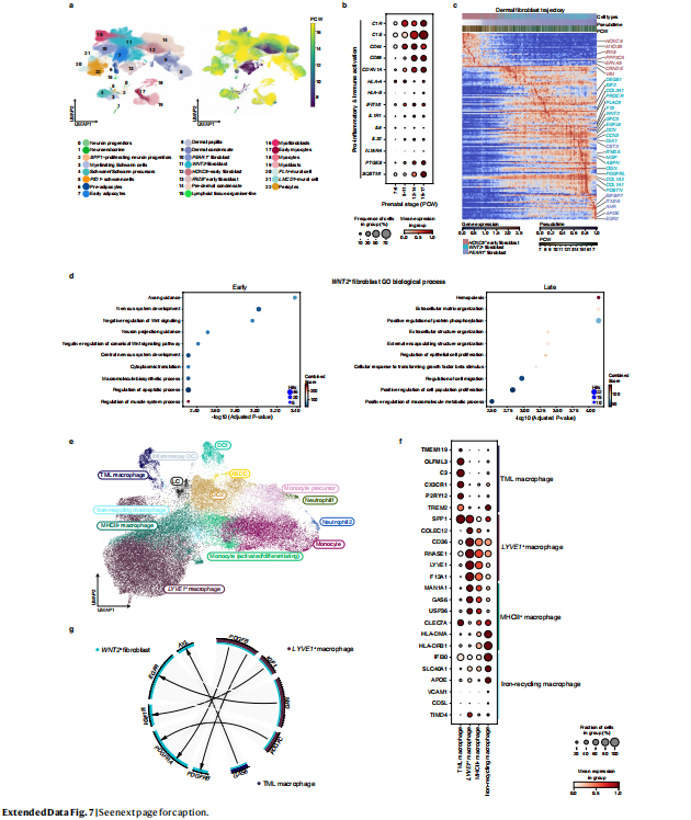
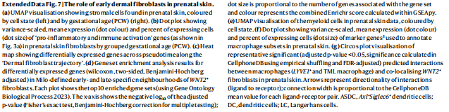
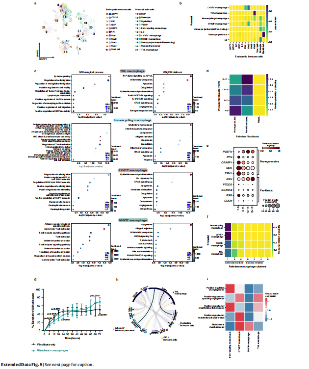
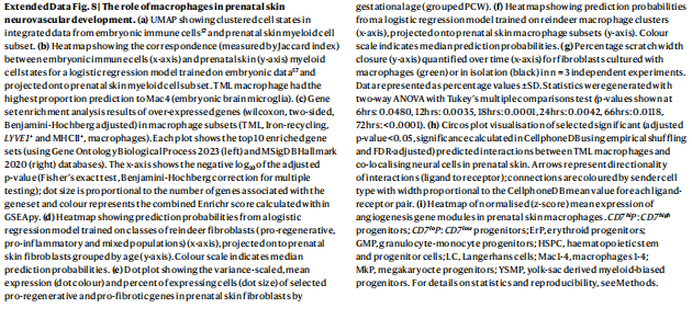
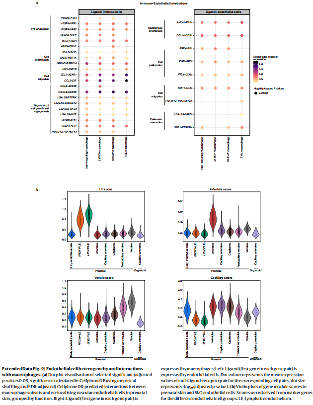
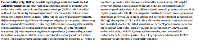
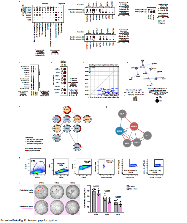
Human prenatal skin is populated by innate immune cells, including macrophages, but whether they act solely in immunity or have additional functions in morphogenesis is unclear. Here we assembled a comprehensive multi-omics reference atlas of prenatal human skin (7–17 post-conception weeks), combining single-cell and spatial transcriptomics data, to characterize the microanatomical tissue niches of the skin. This atlas revealed that crosstalk between non-immune and immune cells underpins the formation of hair follicles, is implicated in scarless wound healing and is crucial for skin angiogenesis. We systematically compared a hair-bearing skin organoid (SkO) model derived from human embryonic stem cells and induced pluripotent stem cells to prenatal and adult skin1 . The SkO model closely recapitulated in vivo skin epidermal and dermal cell types during hair follicle development and expression of genes implicated in the pathogenesis of genetic hair and skin disorders. However, the SkO model lacked immune cells and had markedly reduced endothelial cell heterogeneity and quantity. Our in vivo prenatal skin cell atlas indicated that macrophages and macrophage-derived growth factors have a role in driving endothelial development. Indeed, vascular network remodelling was enhanced following transfer of autologous macrophages derived from induced pluripotent stem cells into SkO cultures. Innate immune cells are therefore key players in skin morphogenesis beyond their conventional role in immunity, a function they achieve through crosstalk with non-immune cells.

Clarisse Ganiera,1 , Pavel Mazinb , Gabriel Herrera-Oropezac , Xinyi Du-Harpura,d, Matthew Blakeleya , Jeyrroy Gabriela , Alexander V. Predeusb ,
Batuhan Cakirb , Martin Preteb , Nasrat Haruna , Jean-Francois Darrigranda , Alexander Haisera , Saranya Wylese , Tanya Shawf , Sarah A. Teichmannb,g,
Muzlifah Haniffab,h,i, Fiona M. Watta,j,1,2 , and Magnus D. Lyncha,k,1,2
Contributed by Fiona M. Watt; received August 6, 2023; accepted November 13, 2023; reviewed by Vladimir Botchkarev and Valerie Horsley
Our understanding of how human skin cells differ according to anatomical site and tumour formation is limited. To address this, we have created a multiscale spatial atlas of healthy skin and basal cell carcinoma (BCC), incorporating in vivo optical coherence tomography, single-cell RNA sequencing, spatial global transcriptional profiling, and in situ sequencing. Computational spatial deconvolution and projection revealed the localisation of distinct cell populations to specific tissue contexts. Although cell populations were conserved between healthy anatomical sites and in BCC, mesenchymal cell populations including fibroblasts and pericytes retained signatures of developmental origin. Spatial profiling and in silico lineage tracing support a hair follicle origin for BCC and demonstrate that cancer-associated fibroblasts are an expansion of a POSTN+ subpopulation associated with hair follicles in healthy skin. RGS5+ pericytes are also expanded in BCC suggesting a role in vascular remodelling. We propose that the identity of mesenchymal cell populations is regulated by signals emanating from adjacent structures and that these signals are repurposed to promote the expansion of skin cancer stroma. The resource we have created is publicly available in an interactive format for the research community.
human cell atlas | skin | basal cell carcinoma | single cell RNA sequencing | fibroblasts
Significance
Single-cell RNA sequencing (scRNAseq) has revolutionised cell biology, enabling highresolution analysis of cell types and states within human tissues. Here, we report a comprehensive spatial atlas of adult human skin across different anatomical sites and basal cell carcinoma (BCC)— the most common form of skin cancer—encompassing in vivo optical coherence tomography, scRNAseq, global spatial transcriptomic profiling, and in situ sequencing. In combination, these modalities have allowed us to assemble a comprehensive nuclearresolution atlas of cellular identity in health and disease.
Author contributions: C.G., F.M.W., and M.D.L. designed research; C.G., X.D.-H., M.B., J.G., and N.H. performed research; C.G., G.H.-O., X.D.-H., A.V.P., B.C., M.P., J.-F.D., A.H., T.S., S.A.T., and M.H. contributed new reagents/ analytic tools; C.G., P.M., G.H.-O., A.V.P., B.C., M.P., A.H., S.W., and M.D.L. analyzed data; and C.G., F.M.W., and M.D.L. wrote the paper.
Reviewers: V.B., Boston University School of Medicine; and V.H., Yale University.
Competing interest statement: In the last 3 y, S.A.T. has been a remunerated Scientific Advisory Board member for GlaxoSmithKline, Qiagen, Foresite Labs, and is a cofounder and equity holder of TransitionBio. F.M.W. and M.D.L. are co-founders of Fibrodyne. F.M.W. and M.D.L. have filed two patents related to skin fibroblasts. Copyright © 2024 the Author(s). Published by PNAS.
This open access article is distributed under Creative Commons Attribution License 4.0 (CC BY).
1 To whom correspondence may be addressed. Email: 该Email地址已收到反垃圾邮件插件保护。要显示它您需要在浏览器中启用JavaScript。, 该Email地址已收到反垃圾邮件插件保护。要显示它您需要在浏览器中启用JavaScript。, or magnus. 该Email地址已收到反垃圾邮件插件保护。要显示它您需要在浏览器中启用JavaScript。.
2 F.M.W. and M.D.L. contributed equally to this work. This article contains supporting information online at https://www.pnas.org/lookup/suppl/doi:10.1073/pnas. 2313326120/-/DCSupplemental.
Published January 2, 2024.

Vitamin D and wound healing: Assessing skin barrier function and implications for chloasma treatment
Qiong Chen1 | Lin Liu2 | Yi Zhang1
1 The Second Affiliated Hospital of Liaoning University of Traditional Chinese Medicine, Shanghai, China
2 Dermatology Department, Jingfu Medical Beauty Hospital of Chunxi Road Ward, Chengdu, China
Correspondence
Yi Zhang, The Second Affiliated Hospital of Liaoning University of Traditional Chinese Medicine, Shanghai, China.
Email: 该Email地址已收到反垃圾邮件插件保护。要显示它您需要在浏览器中启用JavaScript。
This is an open access article under the terms of the Creative Commons Attribution-NonCommercial-NoDerivs License, which permits use and distribution in any medium, provided the original work is properly cited, the use is non-commercial and no modifications or adaptations are made.
© 2024 The Authors. International Wound Journal published by Medicalhelplines.com Inc and John Wiley & Sons Ltd.
Abstract
Chloasma, which is distinguished by irregularities in the pigmentation of skin, poses substantial challenge in the field of dermatology. The regulatory influence of vitamin D on the functions of skin cells implies that it may have the capacity to effectively treat chloasma and promote wound healing. To assess the efficacy of vitamin D in chloasma treatment and its impact on the function of skin barrier during the process of wound healing. The research spanned from April 2022 to September 2023, in Shanghai, China, examined 480 individuals who had been diagnosed with chloasma. A double-blind, placebocontrolled clinical trial was utilized to evaluate effectiveness of topical vitamin D3 in treatment of chloasma. Concurrently, randomized control trial investigated the effects of ingested vitamin D3 supplements on the process of wound healing. Transepidermal water loss (TEWL), chloasma severity score changes, wound size reduction and skin hydration levels were critical performance indiators. Statistically, the severity scores of chloasma decreased significantly in the vitamin D treatment group at 3 and 6 months compared with the placebo (p < 0.05). The Vitamin D group exhibited superior wound healing outcomes, including more substantial reduction in lesion size and enhanced skin barrier function, as evidenced by increased skin hydration and decreased TEWL (p < 0.05). Vitamin D substantially mitigated the severity of chloasma and has beneficial effect on wound healing and integrity of the skin barrier. Based on the results obtained, vitamin D exhibited promise as a therapeutic intervention in the field of dermatology, specifically in treatment of chloasma and promotion of wound recovery.
KEYWORDS
chloasma treatment, dermatology, skin barrier function, vitamin D, wound healing
Key Messages The study aimed to evaluate the effectiveness of vitamin D in treating chloasma and enhancing wound healing.
Findings revealed that vitamin D significantly reduced chloasma severity and improved wound healing, as indicated by increased skin hydration and decreased transepidermal water loss.
