
This article is excerpted from the Clin Exp Dermatol 2024; 49:466–477 by Wound World.
Samuel F. Williams , 1 Helen Wan,1 John Chittock , 1 Kirsty Brown,1 Andrew Wigley,1 Michael J. Cork 1,2,3 and Simon G. Danby 1
1 Sheffield Dermatology Research, Department of Infection, Immunity and Cardiovascular Disease, University of Sheffield Medical School, Sheffield, UK
2 Sheffield Children’s NHS Foundation Trust, Sheffield Children’s Hospital, Western Bank, Sheffield, UK
3 Sheffield Teaching Hospitals NHS Foundation Trust, The Royal Hallamshire Hospital, Sheffield, UK Correspondence: Samuel F. Williams. Email: 该Email地址已收到反垃圾邮件插件保护。要显示它您需要在浏览器中启用JavaScript。
Abstract
Background Atopic dermatitis (AD) is characterized by skin barrier defects that are often measured by biophysical tools that observe the functional properties of the stratum corneum (SC).
Objectives To employ in vivo infrared spectroscopy alongside biophysical measurements to analyse changes in the chemical composition of the SC in relation to AD severity.
Methods We conducted an observational cross-sectional cohort study where attenuated total reflection Fourier transform infrared (ATRFTIR) spectroscopy measurements were collected on the forearm alongside surface pH, capacitance, erythema and transepidermal water loss (TEWL), combined with tape stripping, in a cohort of 75 participants (55 patients with AD stratified by phenotypic severity and 20 healthy controls). Common FLG variant alleles were genotyped.
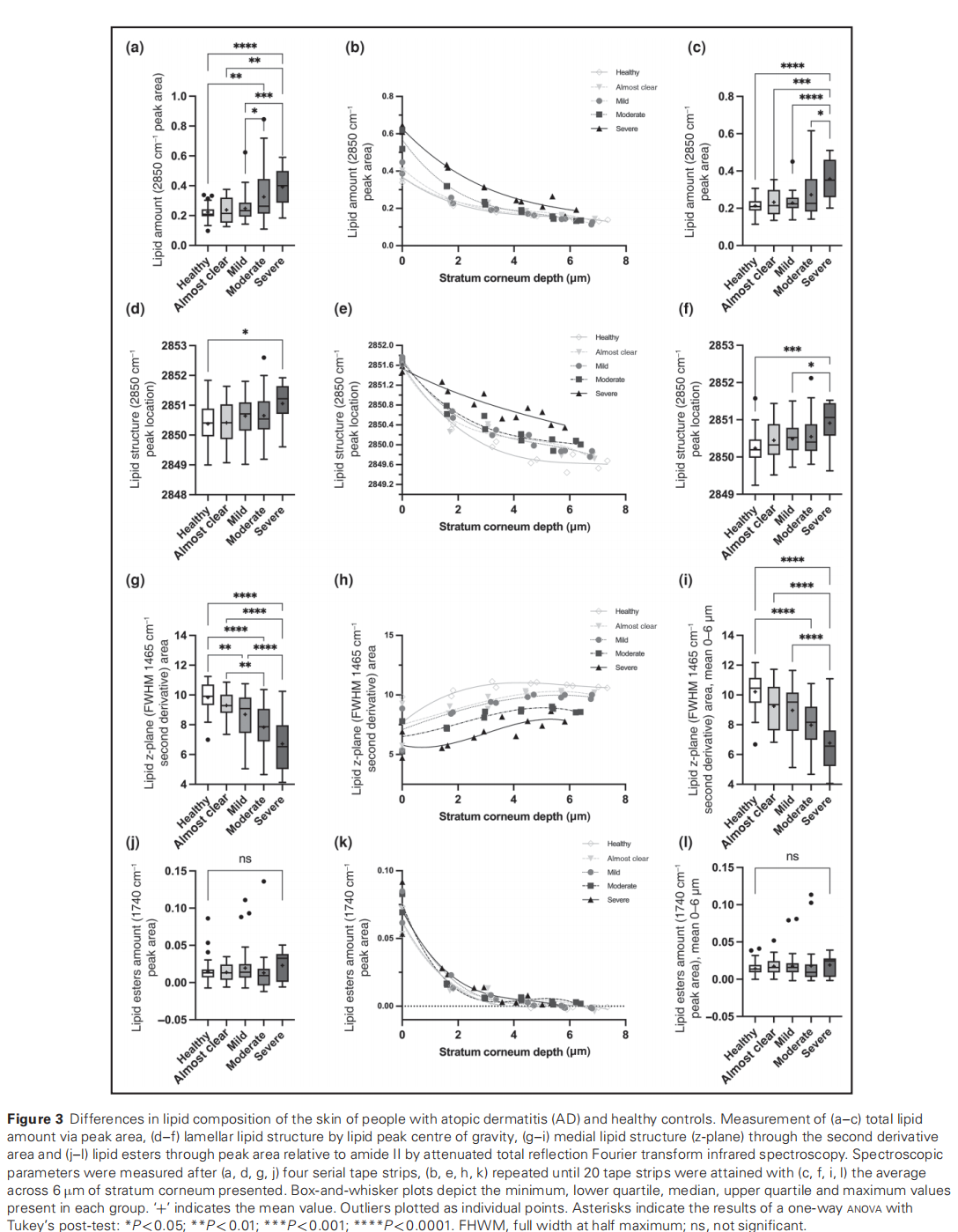
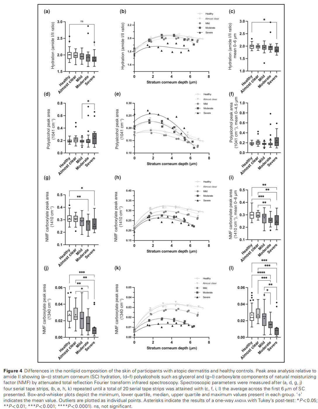
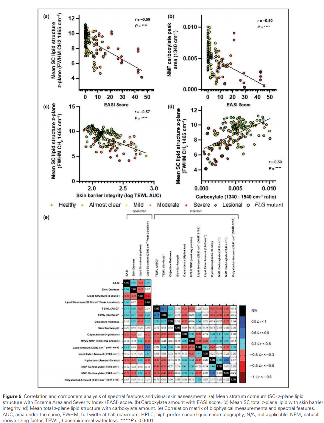
Results Reduced hydration, elevated TEWL and redness were all associated with greater AD severity. Spectral analysis showed a reduction in 1465 cm–1 (full width half maximum) and 1340 cm–1 peak areas, indicative of less orthorhombic lipid ordering and reduced carboxylate functional groups, which correlated with clinical severity (lipid structure r=–0.59, carboxylate peak area r=–0.50).
Conclusions ATR-FTIR spectroscopy is a suitable tool for the characterization of structural skin barrier defects in AD and has potential as a clinical tool for directing individual treatment based on chemical structural deficiencies.
What is already known about this topic?
What does this study add?
Accepted: 20 November 2023
© The Author(s) 2023. Published by Oxford University Press on behalf of British Association of Dermatologists. This is an Open Access article distributed under the terms of the Creative Commons Attribution-NonCommercial License (https://creativecommons.org/licenses/by-nc/4.0/), which permits non-commercial re-use, distribution, and reproduction in any medium, provided the original work is properly cited. For commercial re-use, please contact 该Email地址已收到反垃圾邮件插件保护。要显示它您需要在浏览器中启用JavaScript。


This article is excerpted from the Clin Exp Dermatol 2024; 49:466–477 by Wound World.
