1. Introduction
Deep sternal wound infection (DSWI), also called mediastinitis, is a serious complication after median sternotomy with an incidence of 1% to 5%. While superficial sternal wound infections (SSWI) involve the skin, subcutaneous tissue, and pectoralis fascia only and have much less mortality (0.5% to 9%), DSWI involves retrosternal space, prolongs the hospital stay by an average of 20 days, and is associated with a mortality of 10% to 47% which is double the mortality of those without mediastinitis [1] [2]. The incidence of DSWI is particularly high in the presence of diabetes mellitus (DM), smoking history, chronic obstructive pulmonary disease (COPD), osteoporosis and obesity [1] [2] [3] [4] Prolonged stay in the intensive care unit (ICU), use of assistive devices and reoperation boost the incidence as well. Coronary artery bypass grafting (CABG) is associated with a higher rate of sternal wound infections compared with other surgeries performed through the same surgical approach. Moreover, the technique used in harvesting the internal mammary artery (IMA) for CABG was found to influence the rate of sternal wound infections [5]. When the artery is dissected along the accompanying veins, fascia, adipose tissue and lymphatics (pedicled harvest), the sternal blood flow is decreased by up to 90%, thus increasing the rate of sternal wound infection. In contrast, dissecting the artery free from the surrounding tissues (skeletonized technique) has been shown to preserve the blood supply of the sternum and thereby reduce the rate of sternal wound infections [1]. DSWI is a complication greatly influenced by the surgical technique and can be reduced by a shorter operation and perfusion time and lesser use of electrocautery. On the other hand, shaving with razors, the use of bone wax, reoperation for bleeding, and sternal rewiring are some surgical risk factors that increase the likelihood of this complication [1]. The diagnosis of DSWI could be based on the presence of a group of clinical features such as erythema, fever, drainage and unstable sternum, although a low-grade fever may be the only presentation. According to Pairolero, median sternotomy wound infection could occur within the first week (Type I), or the 2nd to 4th week (Type II) or months to years after surgery (Type III). Most instances of DSWI are of Type II [1]. Unlike SSWI, which is completely resolved with intravenous (IV) antibiotics and local wound care, DSWI is more difficult to cure and requires a much more aggressive treatment regimen. The previous treatment options of DSWI have included closed suction and continuous irrigation while currently, surgical debridement, vacuum-assisted closure (VAC) therapy, flap coverage, and sternal plating are added options [1] [6]. Surgical treatment for angina pectoris was first proposed in 1899. Decades of experimental surgery for coronary artery disease (CAD) finally led to the introduction of CABG in 1964 [7]. Median sternotomy, first proposed by Milton in 1897 [8], was utilized in open heart operation in [3] Iraq for the first time in 1964 by Prof. Yousif D Al-Namaan and his mate Prof. Muayyad M Al-Omeri, cardiothoracic pioneers. Trials of CABG were performed a few years later by Prof. Al-Omeri at the former Republic Hospital (later called Baghdad Medical City), while a modern-time CABG was started by Dr. Najih Al-Asadi (FRCS, a Cardiothoracic & Vascular Surgeon) in 1989 at Al-Rasheed Military Hospital, Baghdad [9]. Despite the seriousness of DSWI after CABG, no study has addressed this problem in our country so far. The current study was conducted in a major tertiary Iraqi cardiac surgical center in order to assess the incidence, clinical and microbial characteristics, perioperative factors, and the outcome of surgical and conservative treatment of this devastating complication in view of the published literature.
2. Patients and Methods
From January 2014 to January 2016, 520 patients underwent CABG in the Iraqi Center for Heart Diseases (ICHD). DSWI was diagnosed in 29 patients (20 males, 69% and 9 females, 31%). Pre-operative, intra-operative and post-operative patient’s risk factors and many variables were studied. A questionnaire form was created. History, patients’ charts (ICU, ward, perfusionist, anesthetic, and surgical records) and admission hospital records of the patients were studied retrospectively; physical examination, diagnosis and DSWI management were studied prospectively. The diagnosis of DSWI was based on the history, physical examination, blood investigations, radiological investigations, and CDC definition criteria for DSWI, which includes the involvement of the deep tissues beyond the skin and subcutaneous tissues, including the fascial, muscle layers, sternum and retrosternal space with or without sternal instability. Patients with DSWI following CABG combined with another procedure and those operated upon elsewhere were excluded from this prospective study. The Ethical Committee of our center approved the study protocol and written informed consent of the patients to participate in the study was obtained.
The patients were thoroughly evaluated. The studied pre-operative variables included age, gender, risk factors such as high body mass index (BMI), DM, COPD and renal impairment, cardiac variables such as LVEF%, number of diseased vessels, type of angina (stable vs. unstable), congestive heart failure, myocardial infarction, previous coronary intervention and pre-operative use of aspirin and low molecular weight heparin (LMWH). Moreover, operative variables such as type of surgery (elective vs. urgent, on-pump vs. off-pump), durations of surgery, cardiopulmonary bypass (CPB) and aortic cross-clamp (ACC), number of grafts and bilateral IMA harvesting were noted. Furthermore, postoperative events such as the length of staying in the ICU and ward, re-exploration for bleeding/tamponade, stroke, coma for >24 hrs and renal impairment were documented. The type of treatment offered to patients with DSWI and death occurring within 30 days of diagnosis of sternal wound infections were reported. All procedures were performed by the same surgical and anesthetic teams. Median sternotomy incision and closure were done according to the standard technique. The number of grafts was dictated by the angiographic and intra-operative findings. Conduits were either the internal mammary artery (IMA) or the great saphenous vein. Most CABG procedures were performed under CPB with topical and central cooling, cross-clamping of the aorta and cardioplegic arrest of the heart, while the off-pump technique was occasionally used. At the end of surgery, the wounds were cleaned with Povidone-Iodine and covered with a 30 cm adhesive gauze plaster, which was kept for 2 days post-operatively.
Parenteral antibiotics (3rd generation cephalosporin and/or penicillin + aminoglycosides) were routinely given for 3 - 5 days post-operatively and then switched to oral antibiotics for 5 days if the patients had an uneventful recovery. Patients with a smooth postoperative course usually stayed for 48 hours in the ICU while those with adverse events stayed longer. The total duration of patients’ stay in the hospital was affected by the presence of the sternal wound infection and other comorbidities such as arrhythmias, myocardial ischemia, renal impairment, cerebrovascular 4 accident, and bleeding. Upon discharge from the hospital, patients prone to sternal wound infection and sternal instability (BMI > 30 kg/m2 , COPD, DM, age > 75 years) received a thoracic vest for 4 - 6 weeks. DSWI was defined according to the guidelines from the US Centers for Disease Control and Prevention (CDC) for post-sternotomy mediastinitis [10], which includes the involvement of the deep tissues beyond the skin and subcutaneous tissues including the fascial, muscle layers, sternum and retrosternal space with or without sternal instability. DSWIs require the presence of one of the following criteria: 1) An organism isolated from a culture of mediastinal tissue or fluid. 2) Evidence of mediastinitis seen during operation; or histopathological examination. 3) At least one of the following symptoms and/or signs with no other recognized cause: chest pain, sternal instability or fever (>38˚C) in combination with either purulent discharge from the mediastinum or an organism isolated from blood culture or culture of mediastinal drainage, or mediastinal widening on chest radiography [1]. Whenever possible, culture verification of the sternal wound infection was attempted by taking wound swabs or wound drainage for culture and sensitivity tests.
The variables of this study were divided into:
1) Demographic.
2) Pre-operative variables include Age, Gender, BMI, DM, HTN, chronic lung disease, renal disease.
3) Pre-operative cardiac variables include LVEF, NO. of the diseased vessels, angina whether stable or unstable, congestive heart failure, myocardial infarction, previous coronary intervention, pre-operative use of anticoagulants, and continuation of aspirin preoperatively. Discontinuation of the anti-platelets was 5 days prior to surgery except for emergency cases.
4) Operative variables including Status of the procedure whether elective or urgent, off or on pump CABG, CPB time, No. of the grafts, bilateral LIMA harvesting, ACC time, and time of surgery.
5) Post-operative variables include ICU stay, ward stay, re-exploration for bleeding/tamponade, stroke, continuous coma for >24 hrs, and renal impairment.
6) Lines of management and early mortality variables including drainage, debridement and wound closure, drainage, debridement and sternal re-wiring, drainage, debridement, sternal re-wiring and closed irrigation, drainage, debridement, resection of the bone, debridement, VAC, steel wire(s) removal, early mortality within 30 days of the diagnosis of the DSWI.
Regarding the surgical technique, the standard approach was median sternotomy. By using a standard pneumatic sternal hand held saw for all of the patients. Patient preparations: The patient lies in a supine position with the arms secured at the sides. The body hair is shaved the night before the procedure, 1 g of 3rd generation cephalosporin or 1 g vancomycin antibiotic prophylaxis is given intravenously within 30 minutes of the incision and the patient is draped according to the institutional protocol with disposable sterile towels covering the skin.
Incision: The incision routinely was done by a median vertical line between the sternal notch and the tip of the xiphoid process with 21 or 22 blade knives. The interclavicular ligament has to be carefully divided followed by digital dissection of the rear surface of the sternum from the underlying sternoclavicular ligament. The xiphoid is severed from the underlying tissue of the diaphragm. The midline is identified by palpating the intercostal spaces and the sternochondral junctions at both sides of the sternum. Osteotomy is performed from above downwards. Bleeding is controlled with pinpoint cautery to avoid continuous blood loss during surgery. The use of bone wax to seal the bone marrow. The number of grafts is decided based on the echocardiographic, angiographic findings of each patient and the intra-operative findings. Conduits used were the harvested internal thoracic artery and long saphenous vein; in 19 patients of the 520 patients bilateral LIMA was used. Off-pump CABG was used in 3 patients out of the 29 and the rest were treated under cardiopulmonary bypass with topical and central cooling, cross-clamping of the aorta and cardioplegic arrest of the heart. Sternal closure: Chest tubes after completion of the cardiac procedure, LIMA bed hemostasis is checked. Mediastinal and/or pleural tubes (28,30F) are placed through stab incisions in the epigastrium. A towel is placed between the heart and the sternal edges for protection, placing the drains below the fascia of the rectus muscle. The stab incisions for the mediastinal drains are made in the epigastrium. Five to eight stainless steel wires are used for closure (either singular or figure of eight). Two wire sutures are placed around the manubrium and four are usually placed around the edges of the body of the sternum. The wires are usually either placed parasternal or through the sternal bone. After all, the wires have been set; the towel is removed carefully while lifting the wires upwards. Before closure, check both retro-sternal halves to rule out bleeding. After proper approximation, the wires are loosely twisted and cut. Then, the ends are twisted further until the sternal halves are tightly re-approximated. The twisted ends must not be too long and usually being buried entirely into the presternal tissue especially in very thin patients. The pectoral fascia is closed with one line of running braided polyglactin absorbable suture followed by a second line of the same type of suture for the subcutaneous tissue. The skin is closed according to the surgeon's preference either with an absorbable or non-absorbable subcuticular running suture or with clips. The skin closure technique was variable. Wound care: Cleaning the wound with iodine and covering it with a 30cm adhesive guaze plaster, which will not be removed until 2 days post-operatively. Sternal care and post-operative period: All the patients with risk for sternal wound infection and instability (body mass index > 30, chronic obstructive lung disease, bilateral mammary harvesting, >75 years of age and diabetes) received a thoracic vest for stabilization for 4 - 6 weeks. I.V antibiotics are routinely kept for 3 - 5 days post-operatively, and then oral antibiotics are to be prescribed for 5 days after discharge from the hospital in the uneventful course of the post-operative period. I.V antibiotics that are used are usually 3rd generation cephalosporins and /or penicillin group antibiotics, with aminoglycosides. The length of ICU stay in average was (48 hrs) for each patient with uneventful post-operative period unless there was an adverse event that required to keep the patient in the ICU. Total hospitalization period post-operatively in average was (seven days) this was affected by complications that necessitated keeping the patient in the hospital, like the following morbidities which includes infection, arrhythmias, myocardial ischemia, renal impairment, cerebro-vascular accident, and bleeding.
Patients of DSWI in this study received one of the following treatments:
1) Drainage, debridement and wound closure;
2) Drainage, debridement and sternal re-wiring;
3) Drainage, debridement, sternal re-wiring and closed irrigation;
4) Drainage, debridement and resection of the bone;
5) Debridement, VAC and steel wire (s) removal.
Each patient with DSWI was followed up for 6 months from the onset of diagnosis of DSWI. Statistical analysis was done using the Excel Sheet of Microsoft office 10. The data were expressed as mean ± SD, ranges, numbers and ratios. P-value was calculated using the chi-square test, Odd’s ratio, and the 2-way contingency table analysis formulas. P value < 0.05 was considered statistically significant.
3. Results
Throughout the study, 520 patients underwent CABG surgery; 425 males (81.7%) and 95 females (18.6%) for varied indications. Twenty nine (29) out of 520 patients developed DSWI diagnosed based on the CDC definition criteria for DSWI, including nine (31.0%) female patients and 20 (69.0%) male patients. Males were significantly more frequently involved than females (p = 0.0001) with a male to female ratio of 20/9 (2.2:1). Male gender was an independent risk factor for DSWI. The age distribution of the patients is shown in Table 1.
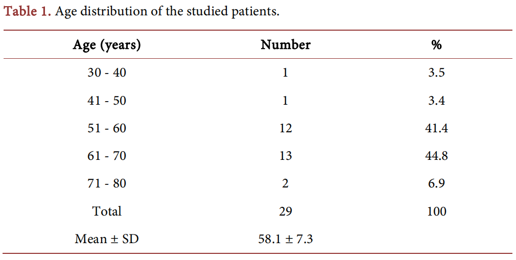
The age of the studied patients ranged between 38 and 75 years, with a mean of 58.1 ± 7.3. Most (n = 25, 86.2%) patients were in the 6th and 7th decades of their lives.
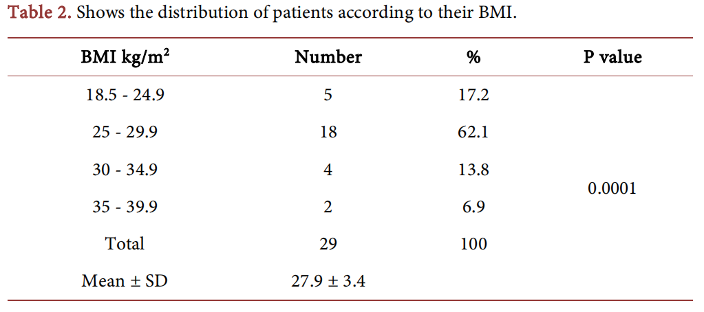
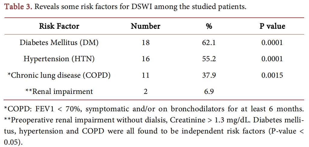
The clinical characteristics and preoperative variables are shown in Tables 2-4. The BMI ranged between 22 and 37.3 with a mean of 27.9 ± 3.4 kg/m2 and it was an independent risk factor for DSWI (P-value = 0.0001). It is worthy to note that only a minority of patients (n = 5, 17.2%) had a normal body weight while the majority (n = 24, 82.8%) were either overweight or obese.
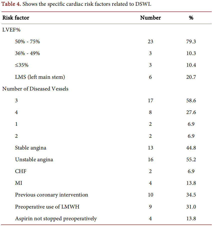
Most patients (n = 23, 79.3%) had a good LVEF% (50-75%). Three to four vessel CAD constituted the majority (n = 25, 86.2%) of cases in this series. The patient had unstable angina more than stable angina (16 vs. 13). Four patients (13.8%) had anti-platelets medication (Acetylsalicylic acid tablets 100 mg) unstopped pre-operatively. Nine patients (31.0%) had pre-operative use of anticoagulants (low molecular weight heparin s.c). A few patients had MI and CHF. The operative characteristics of the patients with DSWI are shown in Table 5.
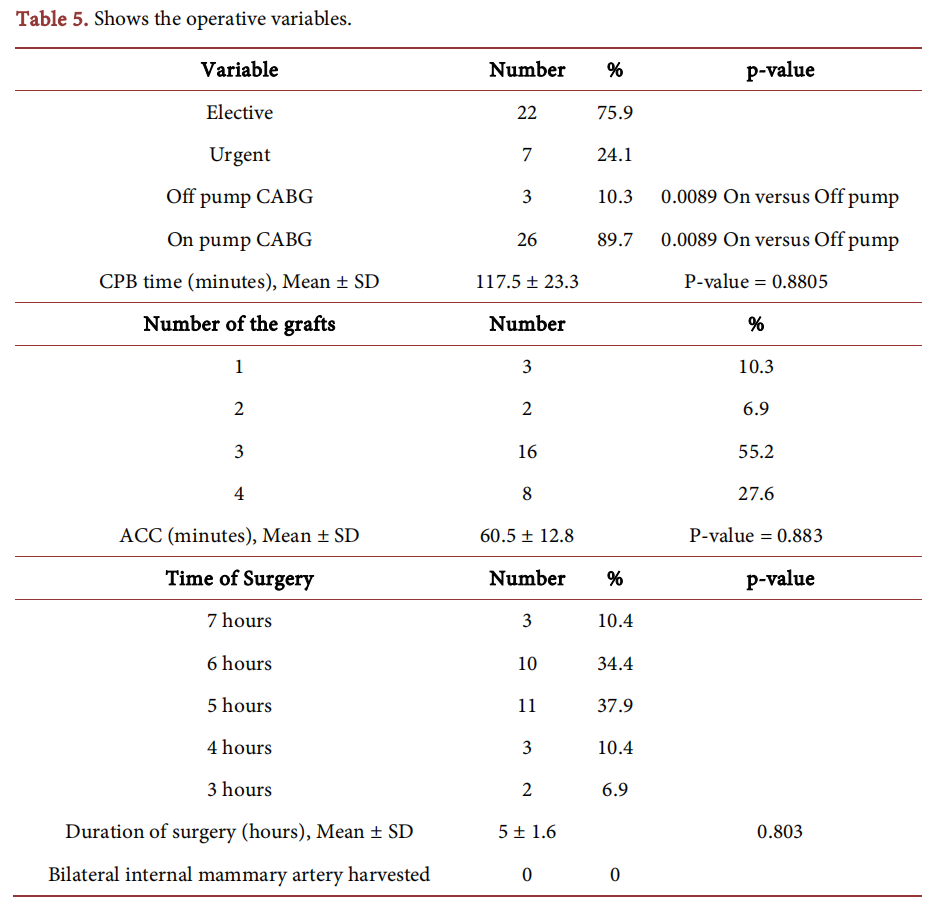
Three-quarters of patients had elective CABG. The majority of procedures (n= 26, 89.7%) were done using CPB and this was an independent risk factor for DSWI (p = 0.0089). Most patients (n = 24, 82.8%) received 3 to 4 grafts. The mean CPB time was 117.5 ± 23.3 minutes and the mean ACC time was 60.5 ± 12.8 minutes while the mean duration of surgery was 5 ± 1.6 hrs. Most surgeries (n = 21, 72.4%) lasted 5-6 hrs. No patient in the study group had bilateral IMA harvesting. Worthy of mentioning that of 520 patients, 19 had bilateral IMA harvesting (3.7%) but none developed DSWI. Relevant postoperative criteria are shown in Table 6.

Renal (n = 8, 27.6%) and neurological complications (n = 5, 17.2%) were on the top. Characteristics of DSWI in the studied patients are shown in Table 7.
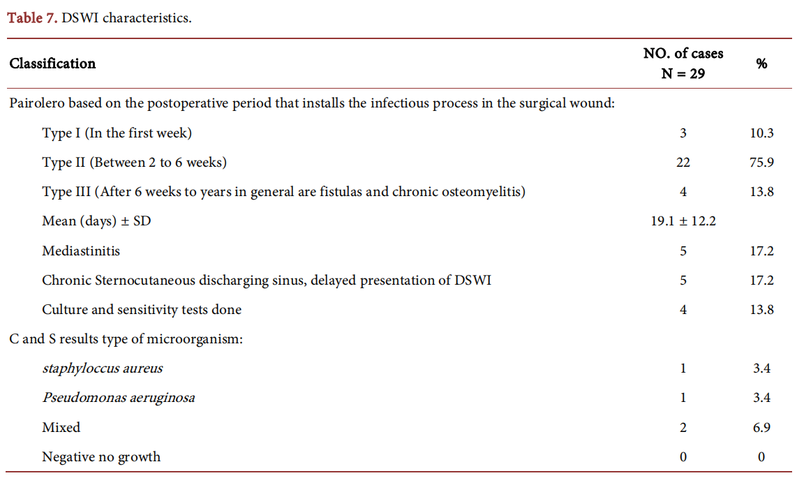
Almost three-quarters of patients had Pairolero Type II DSWI with a mean time of presentation after surgery of 19.1 ± 12.2 days. Only few cases (n = 4,13.8%) were verified by culture which revealed Staphylococcus aureus (n = 1), Pseudomonas aeruginosa (n = 1) and a mixed growth (n = 2). Clinical presentation of the DSWI patients in our study was mostly wound dehiscence with continuous purulent discharge, chest pain, fever, high WBCs count and sternal instability. CXR usually revealed mediastinal widening, slipped or displaced steel wires, and separated sternal edges.
The types of management of DSWI in this study are detailed in Table 8.
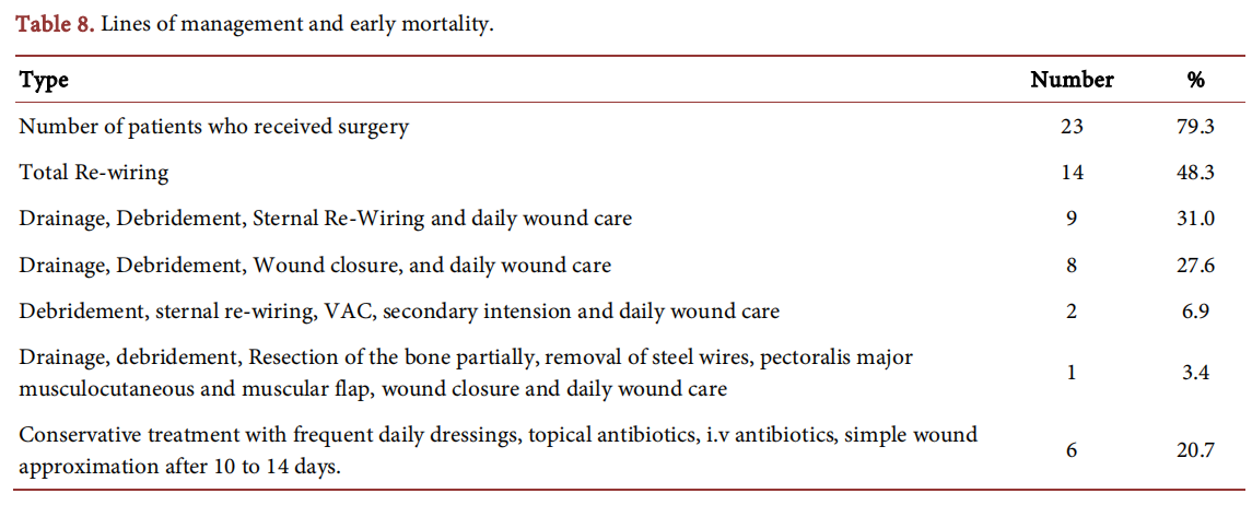
Twenty-three (79.3%) out of 29 patients had been managed surgically, 14(48.3%) of the DSWI patients had sternal re-wiring, meanwhile 2 (6.9%) of DSWI patients had VAC system with other integrated management. Early mortality occurred in 2 (6.9%) patients within 30 days of surgery and both DSWI patients were managed surgically and the cause of death was coma, septicemia and multi-organ failure.
The range of wound healing time was 3 - 24 weeks and the management time of the DSWI patients was uneventful for 18 (62.0%) patients out of the 29 affected.
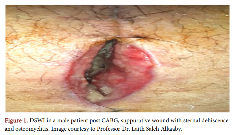
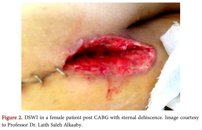
Clinical presentation of the DSWI patients in our study were mostly wound dehescience with continuous purulent discharge, chest pain out of proportion, fever, high WBCs count and sternal instability. By CXR mediastinal wideneing, slipped or displaced steel wires, and space in between the sternal edges, CTscan was done for a few of them to exclude retrosternal collection. Two images were taken from two patients after their approval for showing the general appearance of DSWI (Figure 1 and Figure 2).
Statistically significant variables for DSWI in this study were male gender, high BMI, DM, HTN, COPD and the use of cardiopulmonary bypass.
4. Discussion
Median sternotomy is one of the most commonly used incisions in open heart surgery. DSWI is an uncommon complication, and its improper treatment may result in serious sequelae and even death. Prevention and early recognition of sternal infections are important factors for optimal treatment and management [6]. In this study, we reviewed and followed 520 consecutive CABG patients operated upon in our center from 2014 to 2016. We concluded an incidence of DSWI of 5.6%. Other studies reported lower incidences (1.5% to 1.8%) [2] [11][12]. The relatively high incidence in this study could be attributed to higher rates of uncontrolled hypertension, DM, COPD, and obesity among our patients. Like some other studies, male gender, obesity, DM, hypertension, and COPD were recognized as independent risk factors for DSWIs [6]. Furthermore, Ridderstolpe concluded that another important risk factor for DSWI is the causative microorganism particularly Staphylococcus aureus [6]. Unfortunately, only a few cases of DSWI in the present study were culture-verified as most 9 patients have already received empirical antibiotics at the time of clinical diagnosis. Worthy to note that one of our four cases that had a culture and sensitivity test proved to have a growth of Staphylococcus aureus. The mean age of our patients was 58.1 ± 7.3 years and 25 (86.2%) patients were in the 6th and 7th decades of their lives. Other studies had similar findings [2] [11] [13] and [14]. Old age was identified as a predictor of DSWI by some authors [15] [16] and has been associated with many complications after surgery. Hung Ku reported that with a 1-year increase in patient’s age, the risks of sternal wound infection would be increased by 14%. Cruse and Foord have demonstrated that in patients over the age of 66 years, the chances of developing wound infection are twice as great as in patients between 21 - 50 years of age [16]. The female gender is considered one of the risk factors for DSWI in many scoring systems [13] [17] and [18]. However, male patients were more frequent than female patients with DSWI in our study as well as in some other studies [19] [20].
The mean BMI was 27.9 ± 3.4 kg/m2 and the majority of patients (n = 24,82.8%) were overweight or obese. The preoperative risk stratification of DSWI after coronary surgery considered BMI ≥ 30 kg/m2 as one of the predictors of this complication [17] [18]. Kuduvalli and Birkmeyer found that the risks of DSWI were significantly increased in patients with high BMI. Moulton et al. analyzed 2299 patients after cardiac operations and found that above-average BMI patients were 2.3 times more prone to develop SSWI. Ridderstolpe et al., in a recent analysis of >3000 patients, showed that high BMI patients were 2.1 times more susceptible to sternal wound infections. Likewise, Lu JC et al. found that sternal wound infections were doubled in the patients with this issue. [3]. According to Molina et al., obesity has been identified as the single most important risk factor for postoperative sternal infection in coronary bypass surgery patients. Moreover, being overweight is a major risk factor for sternal dehiscence after any type of cardiac operation with or without infection [4]. The possible reasons for overweight patients having a higher risk for DSWI include the ineffective dose of prophylactic antibiotic, the difficulty of proper skin preparation, adipose tissue providing a good substrate for infection and difficulties in vascular graft harvesting [16]. High BMI was not comparable to Colombier et al. study which didn’t show it as a significant risk factor for DSWI post CABG and only as a dependent risk factor when it is >35. [11] Eighteen (62.1%) patients with DSWI in this series had non-insulin-dependent DM. An equivalent rate was reported by Omran et al. [13]. DM was identified as a predictor of DSWI by many authors [1] [12] [14] [15] and [21]. Hypertension was observed in 16 (55.2%) patients in this series and was comparable to Omran et al. and Kasb et al. [2] [13]. Pre-operative hypertension is a significant risk factor for sternal wound infections, rarely reported in the past [13]. In this series, 11 (37.9%) patients had COPD. Similar rates were reported by other authors [2] [13] [14] [19] while Colombier et al. reported a much lower rate (13.5%) [11]. Patients with COPD are more prone to chest infections and may experience an exacerbation of cough and expectoration in the postoperative period interfering with sternal stability. They may be prescribed steroids beside bronchodilators. The former reduces immunity and increases the chance of wound infection. In the study of Hoseini et al., patients with Grade III and IV NYHA score showed a statistically significant relation with SWI [16]. The critical pre-operative status of the patients undergoing CABG is a predictor of DSWI [18]. Unstable angina (n = 16, 55.2%) among our patients, which was equally reported by other researchers [13] [14], could have contributed to DSWI as well as patients with low LVEF% (n = 6,20.7%). In the present series, 31% of patients didn’t stop their daily 100 mg aspirin and 18.8% of patients continued to use LMWH preoperatively. These medications might have contributed to re-operation for bleeding and thus increased the likelihood of DSWI. Worthy to mention, re-operations for bleeding/tamponade were performed three times (10.3%) in this study which was comparable to other studies [13] [19]. Medalion et al., on the other hand, believe that these medications are not associated with increased postoperative bleeding [22] while Huang et al. found that the risk of re-operation for bleeding was elevated among preoperative aspirin users in patients undergoing valve operations only [23]. Kubota et al. found that when re-exploration for bleeding was performed, mortality was significantly higher than when it was not performed [12]. Cardiopulmonary Bypass was used in 26 (89.7%) patients in this series and was an independent risk factor for DSWI. Hence, the off-pump technique could have been protective. This opinion is shared by Nakano J et al. [21] who found that when off-pump CABG was used for patients with a high risk of DSWI, it showed a significant decrease in the incidence of DSWI. The use of CPB may induce suppression of the immune system and thus predispose to infections [16]. However, our study and some other studies [11] [13] and [19] showed that the time of CPB was not a significant risk factor for DSWI. The duration of the operation is a major risk factor for DSWI [19]. Operations lasting for > two hours are associated with increased infection rates. The longer the duration of surgery, the environmental exposure; hence a higher infection rate is expected [16]. In our series, the mean operative time was 5 ± 1.6 hours and most operations (n = 21, 72.4%) lasted 5 - 6 hours. This could be explained by the severity of CAD in this series [3 - 4 vessels in 25 (86.2%) patients] which required 3 to 4 grafts in 24 (82.8%) patients. An increasing number of grafts were identified by Lu et al. as an independent predictor of DSWI [3]. The use of bilateral internal mammary artery grafts increases the risk of DSWI in patients undergoing CABG surgery [15]. In our series, we didn’t have any patients with bilateral IMA harvesting. But during the study period, 19 of 520 CABG patients had bilateral IMA harvesting without any instance of DSWI. Urgent surgical priority is one of the risk factors for DSWI [17]. In this series, 7 (24.1%) patients had urgent surgeries. The conventional treatment of DSWI usually involves surgical revision followed by open wound dressings with or without wound irrigation. Vacuum-assisted closure has been shown to have several advantages over conventional treatment, including lower in-hospital mortality, improved wound healing and shorter length of stay. Steingrimsson et al. recommend VAC as first-line therapy for most DSWIs following open heart surgery as was adopted in Iceland in 2005 [10]. VAC technique is adopted in wound management to assist in the drainage of necrotic tissue and effusion. This technique is known to increase the capillary diameter and blood flow velocity, as well as to stimulate angiogenesis and endothelial cell proliferation, thereby promoting tissue proliferations and wound closure. These benefits are especially important for seriously infected patients who are unfit for an operation. The VAC technique can be used preoperatively after open wound debridement followed by a secondary reconstruction and flap closure [6]. In the current study, VAC was used twice as an adjuvant to other therapies and the number of treated patients was too small to draw conclusions. Early debridement was advised by Wu et al. study in which primary aggressive management helped in reducing hospital stay, costs, and potentially improve the outcome of the patients with DSWI by preventing it from spreading to more tissues [24]. This aggressive treatment policy is similarly recommended by Shi YD et al. [6]. Similar to other studies [2] [11], most DSWIs in the present series (n = 22,75.9%) were of Type II Pairolero, presenting 2-4 weeks after surgery, and were surgically treated. Apart from re-operation for bleeding which was performed 3 times (10.3%), the top postoperative complication among our patients was acute renal impairment observed in 8 (27.6%) patients. This is an indicator of the seriousness of DSWI as sepsis may affect multiple organs including the kidneys. Unfortunately, we lost two (6.9%) patients because of coma, septicemia and multiple organ failure within one month of surgery. However, this death rate is well below the reported 10% to 47% mortality rate of DSWI [1] [2]. This low mortality could be attributed to early diagnosis of DSWI by the operating surgeon and prompt admission to the hospital once a diagnosis of sternal wound infection is suspected and the initiation of tailored therapy for each patient.
This study does have some limitations. It involved a small number of patients as it was a single-center study; if more cardiac centers were involved, the number would have been higher, and thus, the conclusions would be more valid. Unfortunately, only a few patients were culture verified and the follow-up period was short.
5. Conclusion
DSWI is an uncommon but serious complication of a median sternotomy, particularly following CABG. The meticulous technique of median sternotomy and correction of the modifiable perioperative risk factors is crucial to avoid or minimize the incidence of this complication.
Acknowledgements
My heartfelt appreciation to my mother Asmaa Saleem Esmail Ah-Ghurabi, a Senior Chemist who has provided me with moral and emotional support in my life, and without her support, patience, statistical analysis help and scientific knowledge, this research work would not have been done smoothly. I am also grateful to my father Basil Ismael AlKhateeb and sisters Farah and Basma ALKhateeb who have supported me along the way.
Conflicts of Interest
The authors declare no conflicts of interest regarding the publication of this paper.
References
[1] Singh, K., Anderson, E. and Harper, J.G. (2011) Overview and Management of Sternal Wound Infection. Seminars in Plastic Surgery, 25, 25-33. https://doi.org/10.1055/s-0031-1275168
[2] Kasb, I. and Amr, M. (2016) Management Plan for Deep Sternal Wound Infection Targeting to Survival Free of Recurrence: A Prospective Evaluation Study. Journal of the Egyptian Society of Cardio-Thoracic Surgery, 24, 238-248. https://doi.org/10.1016/j.jescts.2016.04.001
[3] Lu, J.C.Y., Grayson, A.D., Jha, P., Srinivasan, A.K. and Fabri, B.M. (2003) Risk Factors for Sternal Wound Infection and Mid-Term Survival Following Coronary Artery Bypass Surgery. European Journal of Cardio-Thoracic Surgery, 23, 943-949. https://doi.org/10.1016/S1010-7940(03)00137-4
[4] Molina, J.E., Lew, R.S.L. and Hyland, K.J. (2004) Postoperative Sternal Dehiscence in Obese Patients: Incidence and Prevention. The Annals of Thoracic Surgery, 78, 912-917. https://doi.org/10.1016/j.athoracsur.2004.03.038
[5] Berdajs, D., Zünd, G., Turina, M.I. and Genoni, M. (2006) Blood Supply of the Sternum and Its Importance in Internal Thoracic Artery Harvesting. The Annals of Thoracic Surgery, 81, 2155-2159. https://doi.org/10.1016/j.athoracsur.2006.01.020
[6] Shi, Y.D., Qi, F.Z. and Zhang, Y. (2014) Treatment of Sternal Wound Infections after Open-Heart Surgery. Asian Journal of Surgery, 37, 24-29. https://doi.org/10.1016/j.asjsur.2013.07.006
[7] Head, S.J., Kieser, T.M., Falk, V., Huysmans, H.A. and Kappetein, A.P. (2013) Coronary Artery Bypass Grafting: Part 1—The Evolution over the First 50 Years. European Heart Journal, 34, 2862-2872. https://doi.org/10.1093/eurheartj/eht330
[8] Julian, O.C., Lopez-Belio, M., Dye, W.S., Javid, H. and Grove, W.J. (1957) The Median Sternal Incision in Intracardiac Surgery with Extracorporeal Circulation: A General Evaluation of Its Use in Heart Surgery. Surgery, 42, 753-761.
[9] Al-Fattal S. Prof. Yousif Al-Numan [Internet]. Slideshare. 2012 [cited 2021 Nov 11]. https://www.slideshare.net/Ammarko18/ss-11904732
[10] Steingrimsson, S., Gottfredsson, M., Gudmundsdottir, I., Sjögren, J. and Gudbjartsson, T. (2012) Negative-Pressure Wound Therapy for Deep Sternal Wound Infections Reduces the Rate of Surgical Interventions for Early Re-Infections. Interactive CardioVascular and Thoracic Surgery, 15, 406-410. https://doi.org/10.1093/icvts/ivs254
[11] Colombier, S., Kessler, U., Ferrari, E., von Segesser, L.K. and Berdajs, D.A. (2013) Influence of Deep Sternal Wound Infection on Long-Term Survival after Cardiac Surgery. Medical Science Monitor, 19, 668-673. https://doi.org/10.12659/MSM.889191
[12] Kubota, H., Miyata, H., Motomura, N., Ono, M., Takamoto, S., Harii, K., et al. (2013) Deep Sternal Wound Infection after Cardiac Surgery. Journal of Cardiothoracic Surgery, 8, 132. https://doi.org/10.1186/1749-8090-8-132
[13] Omran AS, Karimi A, Ahmadi SH, Davoodi S, Marzban M, Movahedi N, et al. (2007) Superficial and Deep Sternal Wound Infection after More than 9000 Coronary Artery Bypass Graft (CABG): Incidence, Risk Factors and Mortality. BMC Infectious Diseases, 7, Article No. 112. https://doi.org/10.1186/1471-2334-7-112
[14] Lemaignen, A., Birgand, G., Ghodhbane, W., Alkhoder, S., Lolom, I., Belorgey, S., et al. (2015) Sternal Wound Infection after Cardiac Surgery: Incidence and Risk Factors According to Clinical Presentation. Clinical Microbiology and Infection, 21, 674.E11-674.E18. https://doi.org/10.1016/j.cmi.2015.03.025
[15] Tang, G.H.L., Maganti, M., Weisel, R.D. and Borger, M.A. (2004) Prevention and Management of Deep Sternal Wound Infection. Seminars in Thoracic and Cardiovascular Surgery, 16, 62-69. https://doi.org/10.1053/j.semtcvs.2004.01.005
[16] Hoseini, M.J., Naseri, M.H. and Teimoori, M. (2008) Investigation of Deep Sternal Wound Infection after Coronary Artery Bypass Graft and Its Risk Factors. Pakistan Journal of Medical Sciences, 24, 251-256.
[17] Gatti, G., Dell’Angela, L., Barbati, G., Benussi, B., Forti, G., Gabrielli, M., Rauber, E., Luzzati, R., Sinagra, G. and Pappalardo, A. (2016) A Predictive Scoring System for Deep Sternal Wound Infection after Bilateral Internal Thoracic Artery Grafting. European Journal of Cardio-Thoracic Surgery, 49, 910-917. https://doi.org/10.1093/ejcts/ezv208
[18] Biancari, F., Gatti, G., Rosato, S., Mariscalco, G., Pappalardo, A., Onorati, F., et al. (2020) Preoperative Risk Stratification of Deep Sternal Wound Infection after Coronary Surgery. Infection Control & Hospital Epidemiology, 41, 444-451. https://doi.org/10.1017/ice.2019.375
[19] Brunet, F., Brusset, A., Squara, P., Philip, Y., Abry, B., Roy, A., et al. (1996) Risk Factors for Deep Sternal Wound Infection after Sternotomy: A Prospective, Multicenter Study. The Journal of Thoracic and Cardiovascular Surgery, 111, 1200-1207. https://doi.org/10.1016/S0022-5223(96)70222-2
[20] Okonta, K.E., Anbarasu, M., Agarwal, V., Jamesraj, J., Kurian, V.M. and Rajan, S. (2011) Sternal Wound Infection Following Open Heart Surgery: Appraisal of Incidence, Risk Factors, Changing Bacteriologic Pattern and Treatment Outcome. Indian Journal of Thoracic and Cardiovascular Surgery, 27, 28-32. https://doi.org/10.1007/s12055-011-0081-9
[21] Nakano, J., Okabayashi, H., Hanyu, M., Soga, Y., Nomoto, T., Arai, Y., et al. (2008) Risk Factors for Wound Infection after Off-Pump Coronary Artery Bypass Grafting: Should Bilateral Internal Thoracic Arteries Be Harvested in Patients with Diabetes? The Journal of Thoracic and Cardiovascular Surgery, 135, 540-545. https://doi.org/10.1016/j.jtcvs.2007.11.008
[22] Medalion, B., Frenkel, G., Patachenko, P., Hauptman, E., Sasson, L. and Schachner, A. (2003) Preoperative Use of Enoxaparin Is Not a Risk Factor for Postoperative Bleeding after Coronary Artery Bypass Surgery. The Journal of Thoracic and Cardiovascular Surgery, 126, 1875-1879. https://doi.org/10.1016/S0022-5223(03)01324-2
[23] Huang, J., Donneyong, M., Trivedi, J., Barnard, A., Chaney, J., Dotson, A., et al. (2015) Preoperative Aspirin Use and Its Effect on Adverse Events in Patients Undergoing Cardiac Operations. The Annals of Thoracic Surgery, 99, 1975-1981. https://doi.org/10.1016/j.athoracsur.2015.02.032
[24] Wu, L., Chung, K.C., Waljee, J.F., Momoh, A.O., Zhong, L., et al. (2016) A National Study of the Impact of Initial Debridement Timing on Outcomes for Patients with Deep Sternal Wound Infection. Plastic and Reconstructive Surgery, 137, 414e-423e. https://doi.org/10.1097/01.prs.0000475785.14328.b2
This article is excerpted from the World Journal of Cardiovascular Surgery by Wound World.
