Congenital dermal sinuses are a distinctive form of occult dysraphism where patients present with neurological compression, tethering, or meningitis, with an incidence of 1/1500 births (Chatterjee et al, 2020; Venkataramana, 2011). Though certain congenital presentations could initially be subtle, they may have a substantial impact on ambulation. Lower extremity abnormalities are at the front line of such complications. Increased uneven plantar pressure distribution, owing to such deformities, raises the patients’ risk for ulcerations. An open wound is a chief risk factor in the development of osteomyelitis (Anaya et al, 2017).
Children with spina bifida have been reported to have an increased risk of neuropathic foot ulcers (Ebid et al, 2013). Foot and ankle deformities not only result in difficult ambulation and difficult bracing and shoe wear, but also affect cosmetic appearance. They can also result in skin irritation on account of abnormal loading leading to pressure ulcers, and is a primary factor affecting the quality of life in spina bifida sequelae patients.
Methodical physical examination and scientific evaluation are key factors in preparing an appropriate customised surgical treatment plan. In cases of foot and ankle deformity with sensory disorders, the pressure on the weight bearing area is without pain, making the ulcer difficult to heal (Zang et al, 2020). Treating such deformities with complete surgical excision is the current standard of care and is associated with excellent outcomes, predominantly when undertaken at an early stage in the patients’ life (Tubbs et al, 2019).
Here we present a rare case of a young child presenting with chronic foot deformity, secondary to neurological disorders. Such cases, though frequently observed in adults, are seldom noted in the early years. To our knowledge this is a first-of-its-kind case reported in India; the rarity of such a combination of conditions in children inspired us to publish this case. We believe that early diagnosis and treatment of such patients may assist healthcare professionals in the management of such conditions, allowing a better prognosis for this young population.
Case presentation
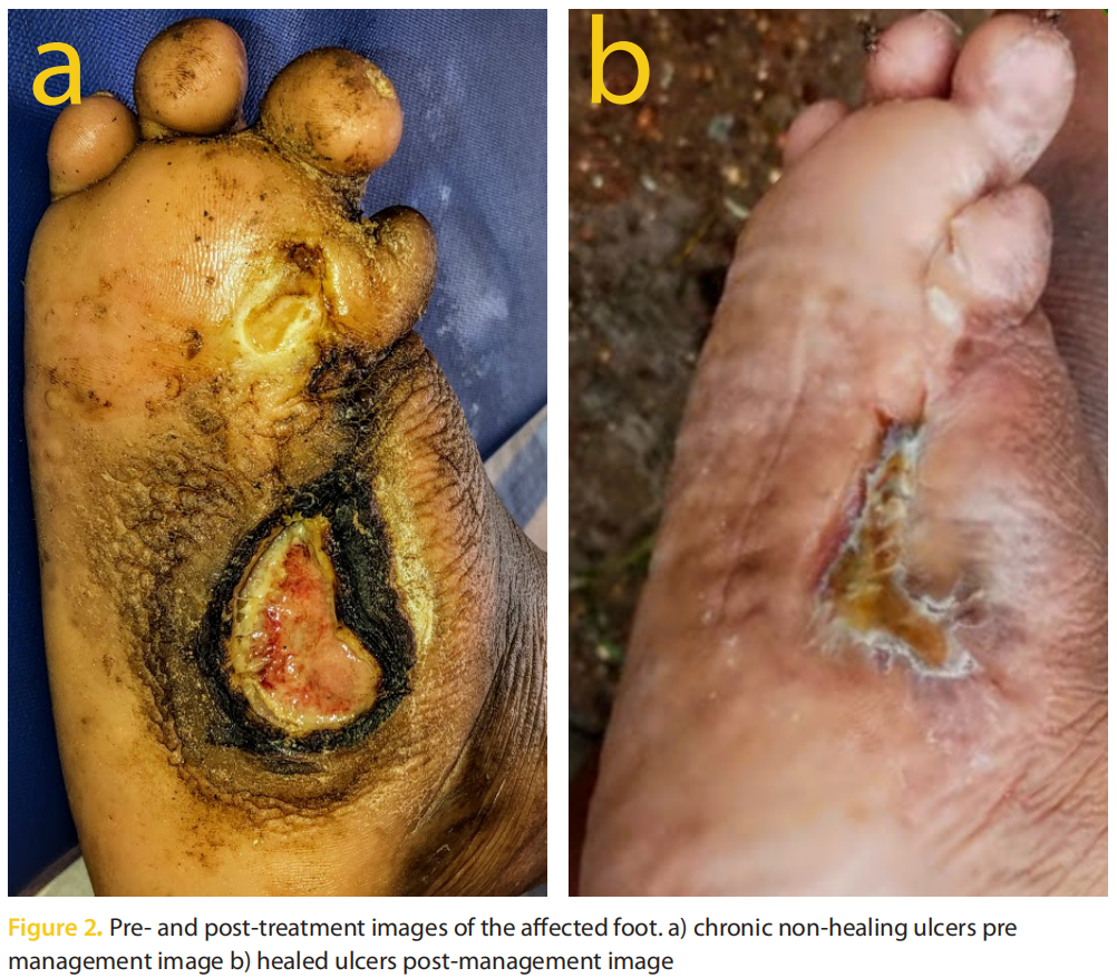
A 12-year-old boy presented to the outpatient department of our clinic with a deformed right foot, swelling in the right foot and leg and multiple chronic non-healing plantar ulcers. Patient history revealed presence of two chronic non-healing plantar ulcers of >6 years duration following a right great toe amputation.
The boy was born with S2–S3 spina bifida that was left unattended by the local health care professional. Despite normal growth, he developed urinary incontinence and difficulty in standing after the age of 1. When the child was 1.5-years-old, he underwent excision of dermal sinus, after which there was worsening of urinary incontinence and gait instability. An ulcer on the right great toe developed 6 months after the dermal sinus excision surgery, which subsequently got infected and was treated by local doctors.
At the age of 4, the child underwent right great toe amputation for the chronic non-healing ulcer and osteomyelitis; the operated wound did not heal. At the age of 5, the boy underwent L1–L5 laminotomy, detethering of the cord, release of adhesions from arachnoiditis and sectioning of filum terminale by a neurosurgeon.
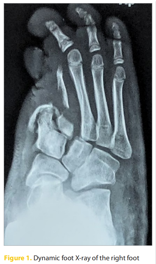
A dynamic right foot X-ray revealed bony deformities and altered biomechanics with altered gait (Figure 1). Neuropathic examination demonstrated that the boy was suffering from sensory neuropathy and the foot was behaving like an insensate foot (no sensation) with pressure ulcers due to altered bony architecture, altered ankle and foot biomechanics.
The child underwent deformity correction surgery with offloading total contact casting (TCC) after obtaining appropriate paediatrician and anaesthetic fitness. Postoperatively, broad spectrum antibiotics were started. Meropenem 500mg IV BD for 3 days. The tissue culture was found to be sensitive to cefoperazone/ sulbactam and tigecycline, after which he was switched over to cepaperazone with salbactum IV BD for 10 days (40 mg/kg/dose). The boy was discharged after 1 week and regular dressing with a bivalved ankle-high removable (offloading) device was planned. Complete healing was achieved within 100 days (Figure 2). Customised footwear for long-term offloading was planned to prevent recurrence and render quality of life.
Discussion
Neuropathy-related ulcerations linked to spina bifida are often slow to heal and unresponsive to treatment, which is further complicated by osteomyelitis. An insensate foot is at higher risk of ulceration and subsequent osteomyelitis. Unless detected early, the prognosis of bone or limb salvage with osteomyelitis is usually very poor. It is therefore imperative to ascertain the association between the neurological deficit and foot deformity in the early years of the patient in order to provide a better prognosis.
A podiatrist is an essential member of a multidisciplinary team that treats patients with neurological disorders due to subclinical podiatric manifestations in neurological disorders. If a podiatrist is consulted early, the progression of the deformity can be delayed. Considering the progressive sequence of events may assist a podiatrist in diagnosing the problem earlier in infancy and aid in taking preemptive measures towards a normal adult life (Anaya et al, 2017). Parents and caregivers also play a fundamental role in the multidisciplinary team (Richterová et al, 2021).
A similar case report of a 44-year-old Caucasian woman was published by Anaya et al (2017). The patient suffered from spina bifida and presented with deformational complications of the foot since childhood that had progressed from multiple lacerations to ulceration and osteomyelitis. She had other side effects of spina bifida including peripheral neuropathy and equinovarus deformity of the left foot, which had resulted in ulcerations.
Another case report of a 44-year-old female with history of a non-healing ulcer of over 20 years was described by Merrill et al (2015). She had a past medical history of spina bifida with peripheral neuropathy and deformity to the left lower extremity. Chronic osteomyelitis in combination with her underlying conditions made closure of her wounds difficult. She underwent a series of surgeries over her lifetime until her wound finally closed.
In 2020, Heidtmann et al reported a 24-year retrospective case series of distal lower extremity manifestations in spina bifida patients of the Yucatan Peninsula, where the most common manifestation was ulcerations. Out of 17 ulcerated limbs, 5 had developed osteomyelitis and 8 were insensate. The first case exemplified a male patient with talipes equinovarus (clubfoot deformity) on the left foot, whose clinical presentation at birth predisposed him to a higher risk of developing clubfoot, given his gender and history of spina bifida. The second case was that of a 2-year-old girl with spina bifida, insensate feet, an active ulcer on the right foot, and flexible bilateral cavovarus deformity. While the third case revealed the most severe result from an insensate lower extremity in spina bifida patients, amputation. This patient was initially seen at the age of 24 years, despite having a rigid cavovarus deformity since birth, resulting in chronic non-healing wounds that eventually led to osteomyelitis. The authors advocated the need for early intervention in any flexible deformity in order to avert them from becoming rigid. They also highlighted that spina bifida patients with concomitant lower extremity deformities ought to seek medical help at a younger age to prevent progression of the deformity and possible limb loss.
Armstrong (2021) asserted that the COVID-19 pandemic and global lockdowns had led to deferred preventative and emergent wound care in patients with chronic wounds. Since limbs and lives are at stake, there is a need for healthcare professionals to manage an influx of such patients with more severe chronic wounds, and those who might require more aggressive treatment approaches.
As our patient was admitted during the pandemic, we faced an array of challenges from the need to reduce hospital stay, cost, and frequency of dressings to early ambulation. Despite these, we accomplished complete healing of the chronic non-healing ulcer with ambulation, in addition to delivering a better quality of life to the adolescent.
One consideration, in such young cases, is that bone ossification occurs only after the age of 16 years. The loss of the middle arch in this case was an imperative loss to the biomechanics of the affected foot. The adolescent will have to live with a lateral arch and appropriate support for the rest of his life. Regular and consistent follow-up in these patients is essential every 2–3 months to assure compliance, and wear and tear should be assessed at least every 4 months depending on activity levels.
Conclusion
Spina bifida, along with numerous other neurological conditions, must be evaluated by a group of multispecialty physicians, together with a podiatrist (wound care professional), for a comprehensive pre-emptive examination. If diagnosed accurately and treated early, many peripheral complications can be evaded and successfully managed without limb-threatening conditions. There must be a multidisciplinary approach to monitor, prevent and treat possible complications that may impact functionality, quality of life as well as survival of the affected individuals.
Declaration of Interest: The author(s) declared no potential conflicts of interest with respect to the research, authorship, and/or publication of this article. The author(s) received no financial support for the research, authorship, and/or publication of this article.
Acknowledgements: We would like to thank Dr. Aafreen Saiyed for providing medical writing services for the article.
References
1. Anaya LAR, Caballes R, Fuentes Y et al (2017) A case report: chronic foot deformities secondary to neurological disorders. Clin Surg 2:1388
2. Armstrong DG (2021) Managing the surge: Delayed chronic wound care during COVID-19. Am J Manag Care https://tinyurl.com/2s39ts4s (accessed 30 March 2022)
3. Chatterjee S, Sil K, Harishchandra LS (2020) Encephalocele, meningocele, and dermal sinus. In: Di Rocco C, Pang D, Rutka J (eds) Textbook of Pediatric Neurosurgery. Springer https://doi.org/10.1007/978-3- 319-72168-2_8 (accessed 30 March 2022)
4. Ebid AA, El-Kafy EMA, Alayat MSM (2013) Effect of pulsed Nd: YAG laser in the treatment of neuropathic foot ulcers in children with spina bifida: a randomized controlled study. Photomed Laser Surg 31(12):565–70. https://doi.org/10.1089/pho.2013.3533
5. Heidtmann A, Madulapally L, Anaya LR, Cawley D (2020) Distal lower extremity manifestations in spina bifida patients of the Yucatan Peninsula: A 24-year retrospective case series. The Foot and Ankle Online Journal 13(4):8. https://doi.org/10.3827/ faoj.2020.1304.0008
6. Merrill TJ, Rodriguez LA, Bernstein M (2015) Spina bifida: a twenty year struggle with osteomyelitis. the podiatry institute update. pp.195-199. https://tinyurl.com/ y37cmjst (accessed 30 March 2022)
7. Richterová R, Kolarovszki B, Opšenák R (2021) Management of Pediatric Patients with Spina Bifida. In: Kolarovszki B, Messina R, Blè V (eds) Spina bifida and craniosynostosis - new perspectives and clinical applications [Internet]. IntechOpen, 2021. https:// tinyurl.com/59s28rex (accessed 30 March 2022)
8. Tubbs RS, Oskouian RJ, Blount JP, Oakes WJ. Occult Spinal Dysraphism. Springer https://doi.org/10.1007/978-3- 030-10994-3 (accessed 30 March 2022)
9. Venkataramana NK (2011) Spinal dysraphism. J Pediatr Neurosci 6(Suppl1): S31–S40. https://dx.doi. org/10.4103%2F1817-1745.85707
10. Zang J, Qin S, Shi L (2020) Lower limb deformity caused by spina bifida sequelae and tethered cord syndrome. In Qin S, Zang J, Jiao S, Pan Q (eds) Lower Limb Deformities. Springer 471–94
This article is excerpted from the Wounds Asia 2022 | Vol 5 Issue 1 by Wound World.

