
伤口世界
- 星期三, 08 7月 2026
次氯酸在伤口护理中的应用研究进展
王昳娜1,何振华1,周瑾1,孙晓芬2
1.绍兴文理学院元培学院,浙江 312000;2.浙江中医药大学附属第二医院
Research progress on the application of hypochlorous acid in wound care
WANG Yi'na,HE Zhenhua,ZHOU Jin,SUN Xiaofen
Shaoxing University,Yuanpei College,Zhejiang 312000 China
Keywords hypochlorous acid solution;wound;infection;bacterial biofilm;clinical application;review
摘要 介绍了次氯酸溶液的发展史、杀菌机制、特点、与其他伤口清洁消毒剂的比较、使用方法以及当前研究的不足,旨在为次氯酸
溶液在伤口治疗、护理中的应用提供依据。
关键词 次氯酸溶液;伤口;感染;细菌生物膜;临床应用;综述

- 星期二, 07 7月 2026
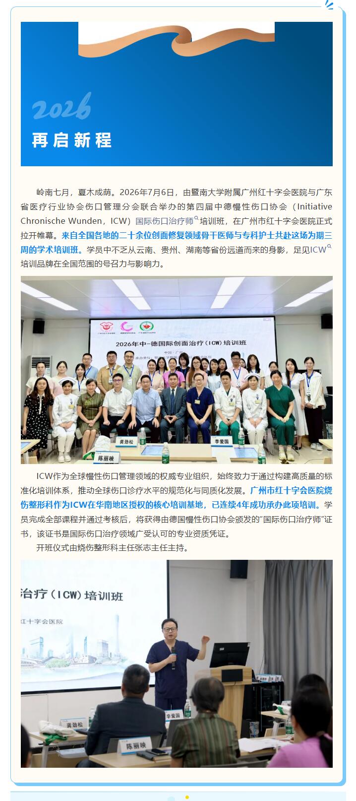
四年深耕,中德携手再谱创面修复新篇章——第四期ICW培训班羊城启幕
本文献转载于红会烧伤,不代表本网站赞同其观点和对其真实性负责,我们只是用于阅读分享,非商业用途,如若侵权,请告知删除。
- 星期一, 06 7月 2026
次氯酸消毒剂的临床应用研究进展
杨金燕1,孙丹1,夏婷婷1,施施1,杨育卉2,闫中强1.3
1.解放军总医院海南医院疾病预防控制科2.血液科,海南 三亚 572014;3.解放军总医院第二医学中心疾病预防控制科,北京 100039
摘要:次氟酸消毒剂以其强大的消毒杀菌能力和环境友好特性,在医疗领域得到广泛关注及应用。有效浓度的次氟酸不仅能高效杀灭细菌、病毒等微生物,同时具有低毒、无残留、低腐蚀性等优势,适用于各种精密设备及复杂的医疗场景。次氯酸消毒剂可用于儿科病房、血液透析室等重点医疗场所的环境消毒。此外,在口腔护理、伤口处理、手术部位消毒等领域展现了独特的优势。本文探讨了次氯酸消毒剂的性能,为其在医疗机构的合理应用提供参考。随着清洁消毒技术的不断进步和应用的深入拓展,次氟酸消毒剂在医疗领域的应用前景将更加广阔。
关键词:次氯酸;次氯酸消毒剂;医疗机构;理化性质;临床消毒
中图分类号:R187 文献标识码:A 文章编号:1005-4529(2025)03-0476-05
Progress of research on clinical application of hypochlorous acid disinfectant
YANG Jinyan*,SUN Dan,XIA Tingting,SHI Shi,YANG Yuhui,YAN Zhongqiang* Hainan Hos pital of PLAGeneral Hospital,Sanya ,Hainan 572014,China
Abstract: Hypochlorous acid disinfectant has been widely noticed and applied in the medical field due to its power-ful disinfecting and sterilizing ability and environmental friendly characteristics.Effective concentration of hypo-chlorous acid disinfectant can not only efficiently kill bacteria,viruses and other microorganisms, but also has theadvantages of low toxicity,no residue and low corrosion,which is suitable for all kinds of complicated equipmentand complex medical scenarios,It can be used for environmental disinfection in major medical places such as pedi-atric wards and hemodialysis rooms,In addition,it demonstrates unique advantages in the oral care,wound treat-ment, and surgical site disinfection, This article explores the properties of hypochlorous acid disinfectant to pro-vide reference for the reasonable application in healthcare facilities,With the continuous progress of cleaning anddisinfection technology and ir-depth expansion of application,the prospect of hypochlorous acid disinfectant appli-cation in the medical field will be broader.
Key words:Hypochlorous acid;Hypochlorous acid disinfectant; Medical institution;Physicochemical property;Disinfection
- 星期六, 04 7月 2026
次氯酸消毒液对皮肤软组织感染常见病原体体外杀菌试验效果分析
高晓东',胡必杰',鲍容3
(1.复旦大学附属中山医院感染管理科2.临床微生物试验室,上海200032)
摘要:目的探讨次氯酸对皮肤软组织感染常见病原体(包括标准茁株及临床茁株)的体外杀菌效果,为临床应用提供依据。方法﹐根抛《消毒技术规范》将0.5左氏单位的临床分离茁株菌悬液与次氯酸作用1 min和10 min,然后将作用后的液体与中和剂混合中和消毒效果,普通病原体的混合液取1.0 ml进行琼脂倾注法培养,化脓性链球菌和非结核分枝杆菌的混合液取0.1 ml分别在血平板和7H10平板上进行涂布法培养;除非结核分枝杆菌培养7d外,其他病原体培莽48h后进行融落计数,计算杀A对数值。结果﹐次氯酸作用标准開株1 min或10min均可将菌量从0.5麦氏单位(相当于107)降剑0;临床菌株包括化脓性链球菌、耐甲氧两林金黄色懒萄球菌(MRSA).耐碳青霉烯类的鲍氏不动朴菌(CRAB).耐碳青霉烯类的铜绿假弟胞菌(CRPA).耐碳青霉烯类的肺炎克需伯杆南(CRKB),脓肿分枝杆南、色分枝杆開和产气荚膜杆開也得到同样结果。结论﹐本试验首次系统全面检测次氛酸对皮肤软组织感染常见的病原体(包括非结核分枝杆開,厌氧掬,真南等)均有很好的体外杀南活性,可以作为感染伤口的局部爪药。
关键词:次氯酸;皮肤软组纠感染;病原体
中图分类号:R187文献标识码:A文章编号:I005 4529(2017)081714 03
Effectiveness of hypochlorous acid solution against common pathogens of skin and soft tissue infection in vitro
GAO Xiao-dong,HU Bi-jie,BAO Rong
(Zhongshan Hospital of Fudan University ,Shanghai 200032,China)
Abstract:ORJECTIVE To investigale the gertmicidal elficientey of hypochlorous acid solution sgainst cotutmotLpathogcns (Inucluding staudeurd strainus anud clinical strains) of skin atnud solt tissuc inlcetion in vitro.so as to provil:the bosis for improving the boctericidal effect.METHODSAccording to " technical standord for disinfection" ,acertain concentrotion of pothogens of clinical isolates wos treated with hypochlorous ocid solution for 1 min or 10 min,anud then the action of the solution anud neutralizer were tmuixeud, 1.0ml of the tuixlure of cotmtmot.puthogens was used lor agar pouring culure,0. 1 ml of the tuixlure ol Sireplococcus pyogenes anud nontuberulosismycobecterie were cultured using the cooting method in the blood plate and 7H10 plate.Unless the nontuberculo-sis mycohscteria was cultured for 7 d, the other pathogens were cultured for 48 h and the colonies were counted.RESULTS Treatment of hypochlorous acid solution for 1 min or 10 min could decrease the amount of bacte-ris from 0.5 Maxwell units (equivalent to 107) to 0.The clinical isolstes of S. pyogenes,methicillin-resistantSta phylococcus aureus (MRSA),carbepenem-resistant Acinetobacter baumannii(CRAR),carbapenem-resistantlP'seudomonas aeruginosa (CRPA),carhapenen-resistant Klebsiella pneumoniae(CRKB),Mycohacterium ab-scess ,Mycobacterium tuberculosis , and Clostridium per fringens also got the sxatme results,CONCLUISION Hypo-chlorous acid solution has good germicidal efficiency against common pethogens of skin and soft tissve infection imvittro,and it can be used as topical treatment for infected wounds.
Key words:Hypochlorous acid solution; Skin and soft tissue infection;Pathogens
- 星期五, 03 7月 2026
次氯酸对大肠埃希菌生物膜的作用及大肠埃希菌感染创面的临床疗效
刘江 1吴宝林 2朱万招 2刘洁 3王彤 4耿毛毛 2白莉 5刘毅 4
1榆 林 市 第 一 医 院 烧 伤 整 形 外 科 ,榆 林 719000;2 宁 夏 医 科 大 学 临 床 医 学 院 ,银 川750000;3 解放军联勤保障部队第 940 医院烧伤整形外科,兰州 730050;4 兰州大学第二医院烧伤整形与创面修复外科,兰州 730030;5 榆林市中医医院重症医学科,榆林719000通信作者:刘毅,Email:该Email地址已收到反垃圾邮件插件保护。要显示它您需要在浏览器中启用JavaScript。
【摘要】目的 探讨次氯酸对大肠埃希菌生物膜的作用及其对大肠埃希菌感染创面的临床疗效。方法 收集从解放军联勤保障部队第 940 医院 5 个临床科室 2019 年 9—12 月 25 例患者(男16 例、女 9 例,年龄 32~67 岁)送检标本分离出的大肠埃希菌菌株中细菌生物膜形成能力最强的 1 株菌进 行 实 验 研 究 。 将 大 肠 埃 希 菌 分 别 与 162.96、81.48、40.74、20.37、10.18、5.09、2.55、1.27、0.64、0.32 μg/mL 的次氯酸共培养,筛选次氯酸最低杀菌浓度(MBC);将大肠埃希菌与筛选的 MBC 次氯酸分别作用 2、5、10、20、30、60 min,筛选次氯酸的最短杀菌时间。分别于培养 6、12、24、48、72、96 h,采用扫描电子显微镜观察大肠埃希菌生物膜形成情况。大肠埃希菌培养 72 h 后,分别加入 1、2、4、8、16 倍MBC的次氯酸,筛选次氯酸对大肠埃希菌的最低生物膜清除浓度(MBEC)。于大肠埃希菌中分别加入 1、2、4、8 倍 MBEC 的次氯酸及无菌生理盐水,作用 10 min 后,采用活/死细菌染色试剂盒检测活、死细胞数,并计算死菌率(样本数为 5)。2020 年 1—12 月,解放军联勤保障部队第 940 医院烧伤整形外科收治 41 例符合入选标准的感染创面患者,对其进行前瞻性随机对照试验。采用随机数字表法将患者分为次氯酸组 21 例[男 13 例、女 8 例,年龄(46±14)岁]和聚维酮碘组 20 例[男 14 例、女 6 例,年龄(45±19)岁]。2 组患者分别用 100 μg/mL 次氯酸、50 mg/mL 聚维酮碘溶液浸透的无菌纱布湿敷,每天换药 1 次。首次换药前、第 10 天换药时,取创面及创缘组织,采用琼脂培养法培养细菌并定量分析组织细菌量。首次换药前和第 3、7、10 天换药时,肉眼观察创面渗出量和肉芽组织生长情况并评分。对数据行单因素方差分析、Dunnett-t检验、独立样本 t检验、Mann-Whitney U 检验、Wilcoxon 符号秩检验、χ2检验或 Fisher 确切概率法检验。结果 次氯酸对大肠埃希菌的 MBC 为 10.18 μg/mL,MBC 的次氯酸对大肠埃希菌的最短杀菌时间为 2 min。培养 6、12 h,大肠埃希菌处于完全游离状态;随着培养时间的延长,大肠埃希菌逐渐聚集、黏附,至培养 72 h 形成成熟的生物膜。次氯酸对大肠埃希菌的MBEC 为 20.36 μg/mL。与 1、2、4、8 倍 MBEC 的次氯酸作用 10 min 后,大肠埃希菌死菌率均明显高于与无菌生理盐水作用 10 min 后(t 值分别为 6.11、25.04、28.90、40.74,P<0.01)。第 10 天换药时,次氯酸组 患 者 创 面 组 织 细 菌 量 为 2.61(2.20,3.30)×104集 落 形 成 单 位(CFU)/g,明 显 少 于 聚 维 酮 碘 组 的4.77(2.18,12.48)×104CFU/g(Z=2.06,P<0.05);次氯酸组和聚维酮碘组患者创面组织细菌量均明显少于首次换药前的 2.97(2.90,3.04)×106、2.97(1.90,7.95)×106 CFU/g(Z 值分别为 4.02、3.92,P<0.01)。第10 天换药时,次氯酸组患者创面渗出量评分明显低于聚维酮碘组(Z=2.07,P<0.05)。与首次换药前比较,次氯酸组患者第 7、10 天换药时创面渗出量评分均明显降低(Z 值分别为−3.99、−4.12,P<0.01),聚106、2.97(1.90,7.95)×106维酮碘组患者第 7、10 天换药时创面渗出量评分均明显降低(Z 值分别为−3.54、−3.93,P<0.01)。第10 天换药时,次氯酸组患者创面肉芽组织生长评分明显高于聚维酮碘组(Z=2.02,P<0.05)。与首次换药前比较,次氯酸组患者第 7、10 天换药时创面肉芽组织生长评分均明显升高(Z 值分别为−3.13、−3.67,P<0.01),聚维酮碘组患者第 7、10 天换药时创面肉芽组织生长评分均明显升高(Z 值分别为−3.12、−3.50,P<0.01)。结论 次氯酸对游离状态和生物膜状态的大肠埃希菌均有杀灭作用,低浓度的次氯酸对成熟的大肠埃希菌生物膜可起到快速杀菌作用,且次氯酸浓度越高,杀菌效果越好。100 μg/mL 次氯酸能有效减少患者大肠埃希菌感染创面的细菌负荷,表现为创面渗出的减少、间接促进肉芽组织生长,较传统外用抗菌剂聚维酮碘疗效更好。
【关键词】 次氯酸; 大肠杆菌; 生物膜; 感染性创面
Effect of hypochloric acid on Escherichia coli biofilm and the clinical efficacy of hypochloric acid for wounds with Escherichia coli infection
Liu Jiang1 , Wu Baolin2 , Zhu Wanzhao2 , Liu Jie3 , Wang Tong4 , Geng Maomao2 , Bai Li5 , Liu Yi4
1Department of Burns and Plastic Surgery, the First Hospital of Yulin, Yulin 719000, China; 2Clinical Medical College, Ningxia Medical University, Yinchuan 750000, China; 3Department of Burns and Plastic Surgery, the 940th Hospital of the Joint Logistic Support Force of PLA, Lanzhou 730050, China; 4Department of Burns andPlastic Surgery & Wound Repair Surgery, Lanzhou University Second Hospital, Lanzhou 730030, China; 5Intensive Care Unit, Traditional Chinese Medicine Hospital of Yulin, Yulin 719000, China Corresponding author: Liu Yi, Email: 该Email地址已收到反垃圾邮件插件保护。要显示它您需要在浏览器中启用JavaScript。
【Abstract】 Objective To investigate the effect of hypochloric acid on Escherichia coli biofilm and the clinical efficacy of hypochloric acid for wounds with Escherichia coli infection. Methods One strain of Escherichia coli with the strongest bacterial biofilm forming ability among the strains isolated from specimens in 25 patients (16 males and 9 females, aged 32−67 years) from five clinical departments of the 940th Hospital of the Joint Logistic Support Force was collected for the experimental study from September to December 2019. The Escherichia coli was cultured with hypochloric acid at 162.96, 81.48, 40.74, 20.37,10.18, 5.09, 2.55, 1.27, 0.64, and 0.32 μg/mL respectively to screen the minimum bactericidal concentration(MBC) of hypochloric acid. The Escherichia coli was cultured with hypochloric acid at the screened MBC for2, 5, 10, 20, 30, and 60 min respectively to screen the shortest bactericidal time of hypochloric acid. The biofilm formation of Escherichia coli was observed by scanning electron microscopy at 6, 12, 24, 48, 72, and 96 h of incubation, respectively. After 72 h of culture, hypochloric acid at 1, 2, 4, 8, and 16 times of MBC was respectively added to Escherichia coli to screen the minimum biofilm eradicate concentration (MBEC) of hypochloric acid against Escherichia coli. After hypochloric acid at 1, 2, 4, and 8 times of MBEC and sterile saline were respectively added to Escherichia coli for 10 min, the live/dead bacterial staining kit was used to detect the number of live and dead cells, with the rate of dead bacteria calculated (the number of samples was 5). From January to December 2020, 41 patients with infectious wounds meeting the inclusion criteria and admitted to the Department of Burns and Plastic Surgery of the 940thHospital of Joint Logistic Support Force of PLA were included into the prospective randomized controlled trial. The patients were divided into hypochloric acid group with 21 patients (13 males and 8 females, aged (46±14) years) and povidone iodine group with 20 patients (14 males and 6 females, aged (45±19) years) according to the random number table. Patients in the 2 groups were respectively dressed with sterile gauze soaked with hypochloric acid of 100 μg/mL and povidone iodine solution of 50 mg/mL with the dressings changed daily. Before the first dressing change and on the 10th day of dressing change, tissue was taken from the wound and margin of the wound for culturing bacteria by agar culture method and quantifying the number of bacteria. The amount of wound exudate and granulation tissue growth were observed visually and scored before the first dressing change and on the 3rd, 7th, and 10th days of dressing change. Data were statistically analyzed with one-way analysis of variance, Dunnett-t test, independent sample t test, Mann-Whitney U test, Wilcoxon signed-rank test, chi-square test, or Fisher's exact probability test. Results The MBC of hypochloric acid against Escherichia coli was 10.18 μg/mL, and the shortest bactericidal time of hypochloric acid with MBC against Escherichia coli was 2 min. Escherichia coli was in a completely free state after 6 and 12 h of culture and gradually aggregated and adhered with the extension of culture time, forming a mature biofilm at 72 h of culture. The MBEC of hypochloric acid against Escherichia coli was 20.36 μg/mL. The Escherichia coli mortality rates after incubation with hypochloric acid at 1, 2, 4, and 8 times of MBEC for 10 min were significantly higher than that after incubation with sterile saline (with t values of 6.11, 25.04, 28.90, and 40.74, respectively, P<0.01). The amount of bacteria in the wound tissue of patients in hypochloric acid group on the 10th day of dressing change was 2.61 (2.20, 3.30)×104colony forming unit (CFU)/g, significantly less than 4.77 (2.18, 12.48)×10
4CFU/g in povidone iodine group (Z=2.06, P<0.05). The amounts of bacteria in the wound tissue of patients in hypochloric acid group and povidone iodine group on the 10th day of dressing change were significantly less than 2.97 (2.90, 3.04)×106 and 2.97 (1.90, 7.95)×106 CFU/g before the first dressing change (with Z values of 4.02 and 3.92, respectively, P<0.01). The score of wound exudate amount of patients in hypochloric acid group on the 10th day of dressing change was significantly lower than that in povidone iodine group (Z=2.07, P<0.05). Compared with those before the first dressing change, the scores of wound exudate amount of patients in hypochloric acid group on the 7th and 10th days of dressing change were significantly decreased (with Z values of −3.99 and −4.12, respectively, P<0.01), and the scores of wound exudate amount of patients in povidone iodine group on the 7thand 10th days of dressing change were significantly decreased (with Z values of −3.54 and −3.93, respectively, P<0.01). The score of wound granulation tissue growth of patients in hypochloric acid group on the 10th day of dressing change was significantly higher than that in povidone iodine group (Z=2.02, P<0.05). Compared with those before the first dressing change, the scores of wound granulation tissue growth of patients in hypochloric acid group on the 7thand 10th days of dressing change were significantly increased (with Z values of − 3.13 and − 3.67, respectively, P<0.01), and the scores of wound granulation tissue growth of patients in povidone iodine group on the 7th and 10th days of dressing change were significantly increased (with Z values of −3.12 and −3.50, respectively, P<0.01). Conclusions Hypochloric acid can kill Escherichia coli both in free and biofilm status. Hypochloric acid at a low concentration shows a rapid bactericidal effect on mature Escherichia coli biofilm, and the higher the concentration of hypochloric acid, the better the bactericidal effect. The hypochloric acid of 100 μg/mL is effective in reducing the bacterial load on wounds with Escherichia coli infection in patients, as evidenced by a reduction in wound exudate and indirect promotion of granulation tissue growth, which is more effective than povidone iodine, the traditional topical antimicrobial agent.
【Key words】 Hypochloric acid; Escherichia coli; Biofilm; Infectious wounds

- 星期四, 02 7月 2026
弱酸性次氯酸消毒液杀菌性能和腐蚀性的实验研究
李志钢(帝森福德(北京)国际环境科技有限公司,北京 100020)
摘要:弱酸性次氯酸 (HClO) 消毒液作为一种常见的消毒液被广泛应用。文章主要分析了弱酸性 HClO 消毒液的杀菌性能和腐蚀性,采用了理化分析方法和载体定量杀菌实验方法。进行的实验包括中和剂鉴定实验、载体浸泡定量杀菌实验
和金属腐蚀性实验。实验表明弱酸性 HClO 消毒液的杀菌效果较好,腐蚀性较弱。
关键词:弱酸性次氯酸消毒液;杀菌性能;腐蚀性
中图分类号:TQ421
文献标志码:A
文章编号:1008-4800(2023)03-0033-03
DOI:10.19900/j.cnki.ISSN1008-4800.2023.03.010
Experimental Study on Bactericidal Property and
Corrosivity of Weak Acid Hypochlorite Disinfectant
LI Zhi-gang (Dienford (Beijing) International Environmental Technology Co., Ltd., Beijing 100020, China)
Abstract: Weak acid hypochlorite (HClO) disinfectant is widely used as a common disinfectant. This paper mainly analyzes the germicidal performance and corrosivity of weak acid HClO disinfectant, and adopts the physical and chemical analysis methods and carrier quantitative germicidal test methods. The tests carried out include neutralizer identification test, carrier immersion quantitative germicidal test and metal corrosion test. The test shows that the weak acid HClO disinfectant has good germicidal efficacy and weak corrosivity.
Keywords: weak acid hypochlorite disinfectant; bactericidal property; corrosivity
